Journal of Neurology, Neurological Science and Disorders
Reference Electrode Placement in First Dorsal Interosseous (FDI) Muscle during Ulnar Nerve Conduction Studies: A Comparative Evaluation
Chief Clinical Physiologist (Neurophysiology), Neurophysiology Department, Ysbyty Gwynedd Hospital, Bangor, North Wales, LL57 2PW, UK
Author and article information
Cite this as
Hirani S. Reference Electrode Placement in First Dorsal Interosseous (FDI) Muscle during Ulnar Nerve Conduction Studies: A Comparative Evaluation. J Neurol Neurol Sci Disord. 2025; 11(1): 001-006. Available from: 10.17352/jnnsd.000064
Copyright License
© 2025 Hirani S. This is an open-access article distributed under the terms of the Creative Commons Attribution License, which permits unrestricted use, distribution, and reproduction in any medium, provided the original author and source are credited.When the Compound Muscle Action Potential (CMAP) is recorded in motor nerve conduction studies, the reference (E2) electrode can make a significant contribution to the CMAP. Recording from FDI muscles presents a challenge in placing the reference electrode due to its positive deflection. This study investigates the E2 recorded signal and its effect on CMAP measurements when E2 electrode is placed at different sites.
The aim of this research is to determine the optimal position for placing the reference electrode in FDI muscles.
Method: A total of 46 hands were included in this study. Data collection adhered to the extensive and detailed descriptions provided in various research papers. The tests were conducted by a qualified clinical physiologist specializing in Neurophysiology, utilizing a Keypoint 9033A07 machine, in accordance with the Ulnar Nerve Screening Protocol (Protocol 1.1, 2020). All data were recorded numerically to ensure methodological reliability. The CMAP was recorded using the active electrode on the muscle belly and 5 different E2 electrodes placed at distal sites, including the tendon of the FDI muscles at the base of digit II, over the thumb, the tendon of ADM muscles at the base of digit V, Radial pathways at the wrist and tendon for FDI muscles in other hand.
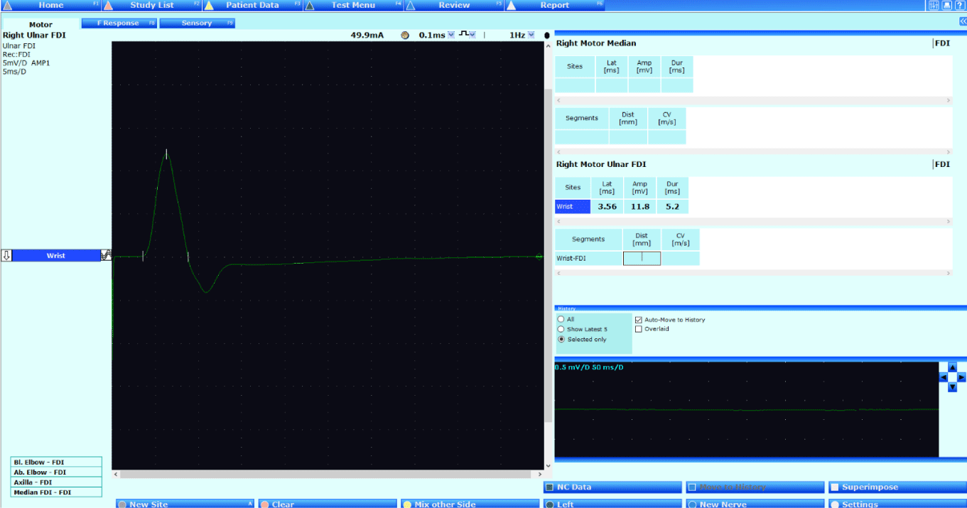
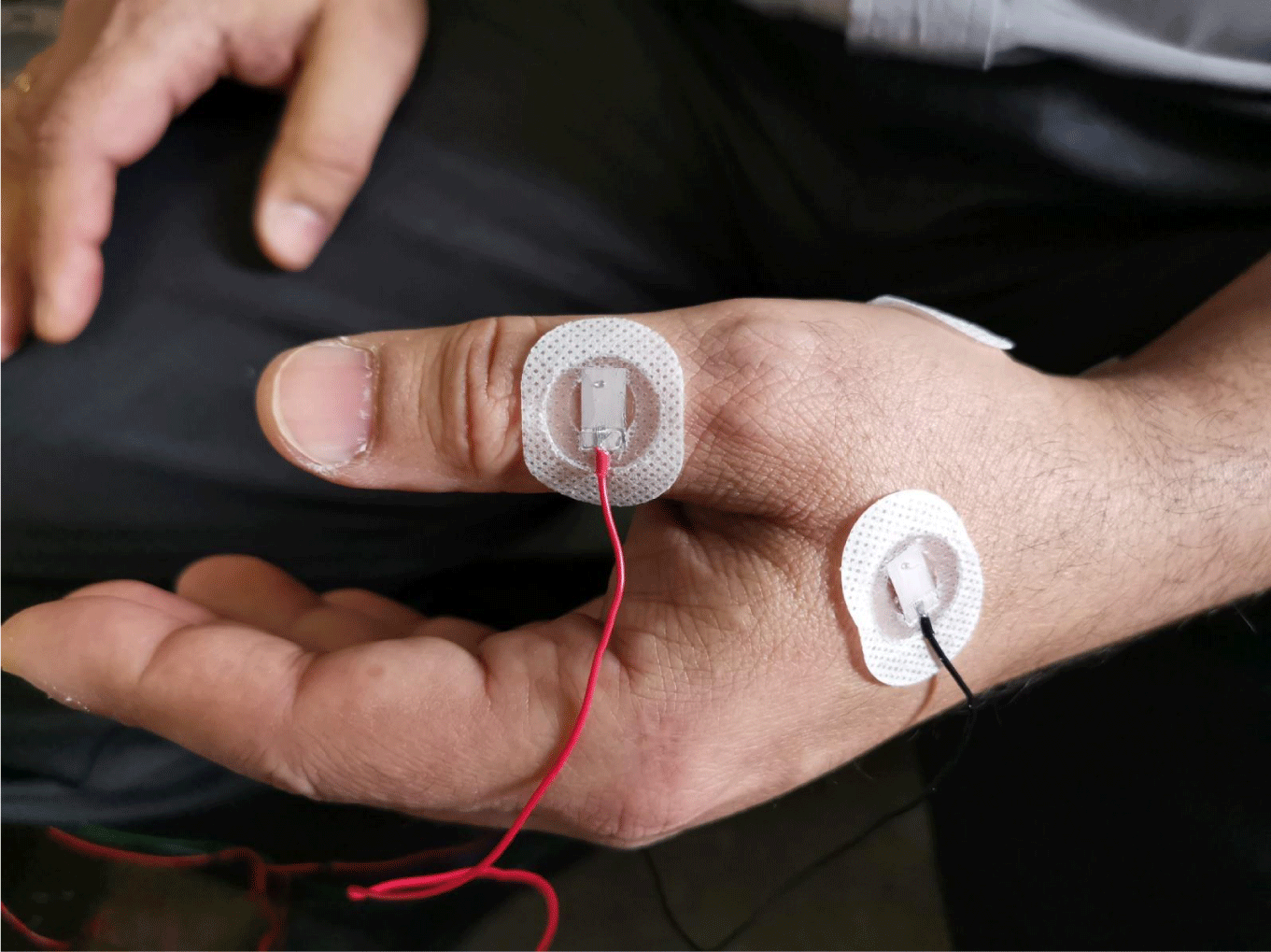
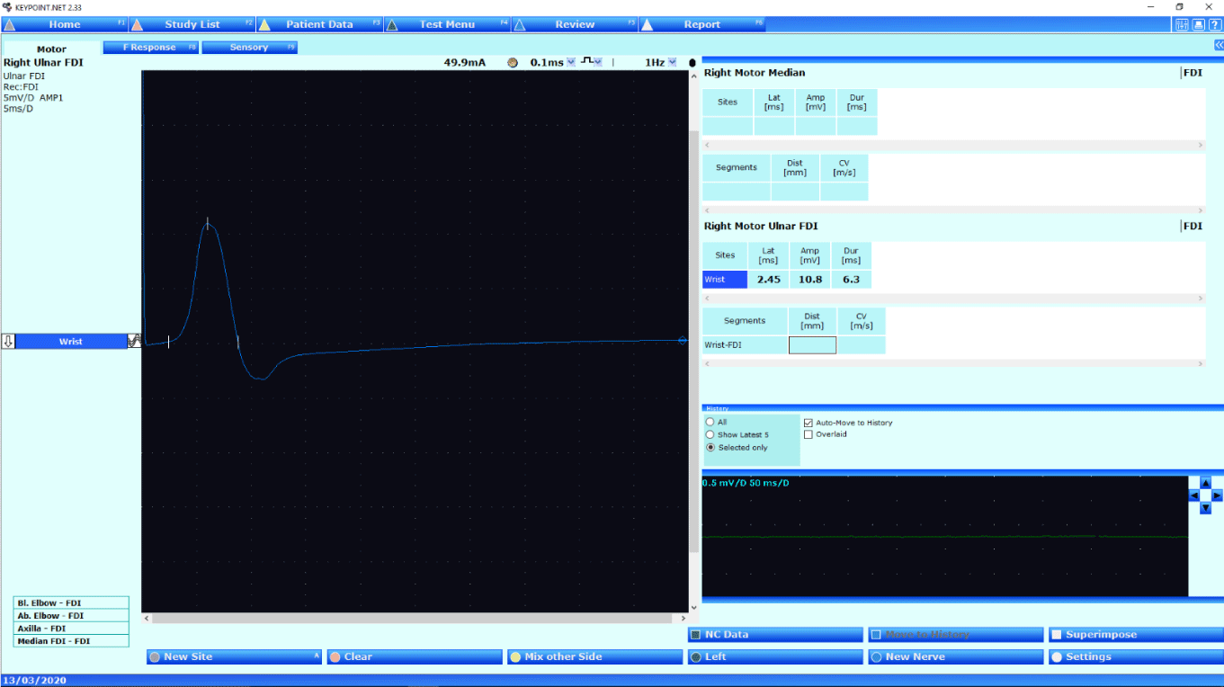
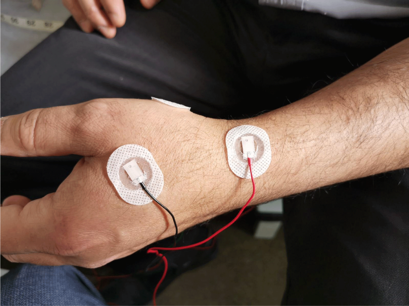
Result: Out of 46 hands were tested for the Nerve Conduction Study (NCS) by placing reference electrodes in five different places while recording ulnar nerves from FDI muscles i.e. Tendon of the FDI muscles to the base of digit II shows positive deflection in all hands with amplitudes ranging from 6 to 15 mV, over the middle of the thumb shows the baseline slightly elevated, impacting distal motor latency calculations with amplitude between 5-8mV, tendon of ADM muscles at the base of digit V shows clear baseline for accurate distal motor latency with the higher amplitude rage 10-18mV, radial pathways of the wrist shows slightly elevated distal motor latency with the amplitude range between 5-10mV and to record from tendon of the FDI muscles in other hand placing the reference electrode shows no clear baseline distal latency with the amplitude range between 5-10 mV.
Conclusion: This study shows that recording the best and clearest response by placing the reference electrode at the tendon of ADM muscles at the base of digit V while recording from FDI muscles of the ulnar nerve is more reliable compared to other four areas to get the maximum amplitude. It also shows that distal motor latency in all placements is comparable.
There are a few different definitions available in books, the internet and in research papers to understand the reason for using a reference electrode (E2) with an active electrode (E1) and the ground electrode (E0). According to Wikipedia, “A reference electrode is an electrode that has a stable and well-known electrode potential. The overall chemical reaction taking place in a cell is made up of two independent half-reactions, which describe chemical changes in the two electrodes. To focus on the reaction at the working electrode, the reference electrode is standardized with constant (buffered or saturated) concentrations of each participant of the redox reaction. According to Pavithara (online source), the reference electrode is defined as, the definition of a reference electrode is: “The electrode potential of any other electrode that can be measured is called a reference electrode. In other words, the electrode whose half-cell potential is known and is constant and completely insensitive to the composition of the solution is called a reference electrode. The reference electrode can act as both anode or cathode, depending upon the nature of other electrodes. As noted by Sanjeev Nandedkar, “The E2 (or “reference”) electrode is conventionally placed in a supposedly electrically quiet or “inactive” location. In most instances, this is at the distal muscle tendon or even further distally. Hence, the CMAP recording is thought to primarily represent the electrical activity of the muscle over which the E1 electrode is placed, and the E2 is considered to be “minimally contributory to the signal”. And, in the words of Jun Kimura in Electrodiagnosis in disease of the Nerve and Muscles principle and practice Fourth edition, page number 1063, he mentioned “Reference electrode is synonymous with input terminal 2 or E2, which remains neutral. “This results in an upward deflection of the impedance.” In most electrochemical measurements, it is necessary to keep one of the electrodes in an electrochemical cell at a constant potential. This so-called reference electrode allows control of the potential of a working electrode. In the past, the electrodes were called ‘G1′, ‘G2′ or E1, E2 and ‘ground’ or G. The terms G1 and G2 refer to the grids of vacuum tubes used in old amplifiers which are no longer available. Later, these inputs were referred to as ‘active’, ‘reference’ and ‘ground’. The term ‘ground’ is confusing. The ‘ground’ in Electrodiagnostic recording refers to a point on the amplifier circuit that is used as a point of reference for voltage measurement. The ‘reference’ electrode is presumed to be electrically silent but does record large volume-conducted potentials. In current practice, the E2 electrode records significant voltage from other muscles innervated by the stimulated nerve [1-8]. The objective of this study is to investigate the effect of the E2 position on the CMAP waveform and its measurements. By reducing E2 contribution, the CMAP will better represent the electrical activity of the tested muscle, both in normal muscles and muscles affected by neuromuscular disorders [9,10]. Ulnar nerve entrapment is the second most frequent neuropathy in the hand, occurring at the wrist and across the elbow Preston and Shapiro suggested that ulnar motor nerve conduction study with FDI recording should be done in all patients with suspected UNW.(define) There are various techniques that have been developed to diagnose the entrapment. The two main muscles at the wrist, the first dorsal interosseous (FDI) and Abductor Digiti Minimi (ADM) are supplied by the deep branch of the Ulnar nerve. Many studies indicate that the FDI muscle can be used to diagnose early entrapment across the elbow. When recording from FDI muscles, there is a challenge in placing the reference electrode at the belly of the FDI muscle at the base of digit II due to its positive deflection.
FDI recording has a shortcoming: the initial positive deflection of Compound Muscle Action Potential (CMAP), which complicates measuring distal motor onset latency. In 1985, Olney and Wilbourn noted that placing Active (E1) over the motor point often did not yield the largest-amplitude response. Dumitru and King demonstrated in 1991 that both a muscle’s origin and its insertion hand produce monophasic potentials. In 1993, Kincaid compared CMAPs obtained with a belly-tendon montage to those recorded over the muscle or tendon with a contralateral reference electrode. The Association of Neurophysiological Scientists (ANS) and British Society of Clinical Neurophysiology (BSCN) published joint guidelines in a BSCN-EPTA Statement on Handheld Devices for Carpal Tunnel Testing in 2007. Citing to their guidelines, Guideline 5 suggests performing motor nerve conduction in the ulnar nerve of the affected limb with surface electrodes to measure response amplitude and latency/velocity. However, it lacks specific instructions on which muscles to record from. The Joint ANS/BSCN Ulnar Nerve Audit 2014, Standard 4, states that ulnar motor nerve conduction should be tested in the affected hand with stimulation points just proximal to the wrist and both proximal and distal to the elbow. This standard also does not specify electrode placement or recording muscles [11-15].
Currently, Neurophysiology services in the United Kingdom lack a unified standard. Each clinic follows its own departmental guidelines.
No clinical assessments will be conducted for the purposes of this research during the Neurophysiological test, to eliminate bias in the patient’s condition.
Method
No ethical approval and institutional approval were needed as this was a retrospective data collection.
Verbal consent was obtained from all participants from each patient prior to using their numerical nerve conduction data for the research purpose. No patient personal identifiable data was included in this study. All patients included in this study had normal nerve conduction studies. This supplementary analysis aimed to establish normative amplitude and latency values. No proximal nerve conduction study data was included in this study as it was not part of the research.
To establish a standard, published literature was reviewed and normative data collected. A Clinical Physiologist conducted the test using a Keypoint 9033A07 machine, following the Ulnar nerve screening protocol (2020). Quantitative data collection ensured accuracy and eliminated bias, including only normal patients, to prevent misinterpretation.
The procedure began by carrying out the sensory testing, by placing the ring electrodes on digit V to stimulate the ulnar nerve and the recording electrode on the surface of the wrist to the allocated nerve. The orthodromic technique was used for both sensory and motor NCS tests. Skin temperature was maintained above 30 °C throughout the procedure throughout the recording. Proper skin preparation was done prior to placing the electrodes. A supramaximal current is applied to obtain the full evoked response from the ulnar nerve at digit V for ulnar sensory recording and motor ulnar nerve pathways from the First Dorsal Interosseous (FDI) of the wrist. Amplitude was recorded from peak to peak for sensory responses, and base to peak for motor responses.
All patient data was collected by fulfilling the criteria mentioned in the above paragraph. The criteria mentioned in the above paragraph are intended to be more reliable from a Clinical Physiologist’s perspective.
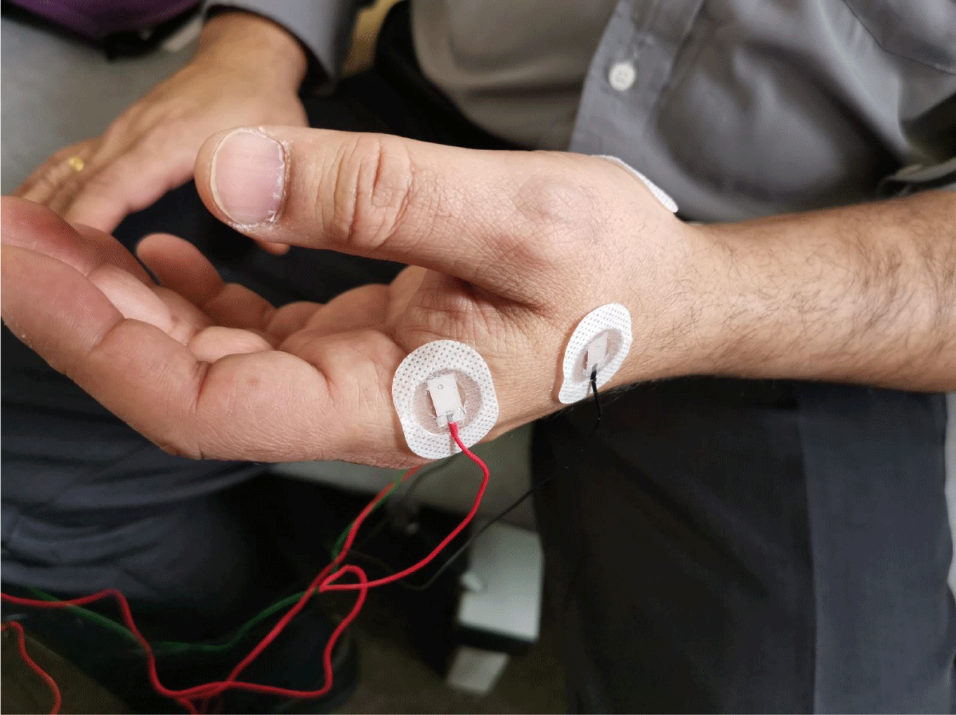
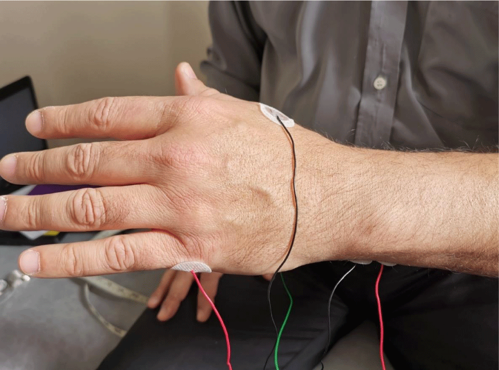
The reference electrode in motor ulnar nerve conduction study, while recording from FDI muscles were placed at five sites:
- The active electrode at the belly of FDI muscles,
- The ground electrode between stimulation and recording sites,
- The reference electrode over various points:
- Base of digit II (tendon of FDI muscles)
- Middle of the thumb
- Base of digit V (tendon of ADM muscle)
- Contralateral hand FDI muscle
- Radial nerve pathways at wrist.
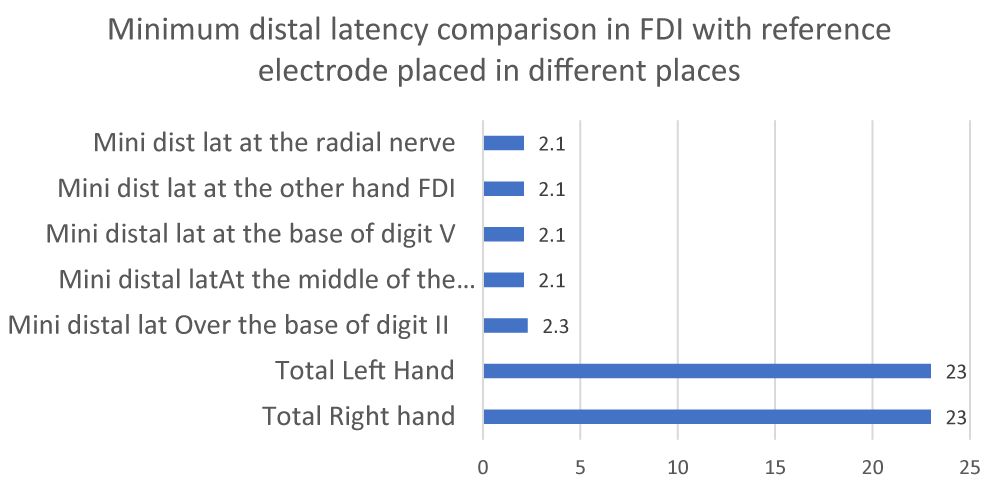
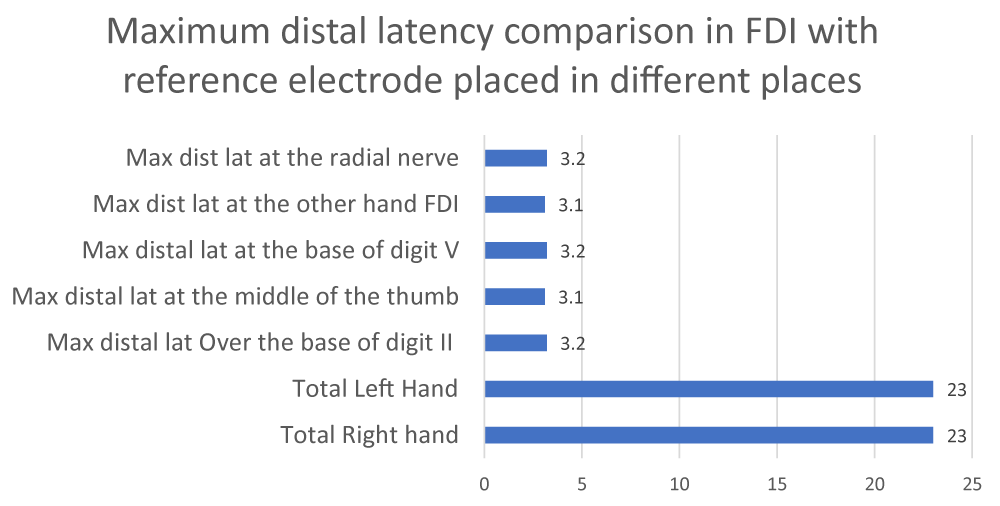
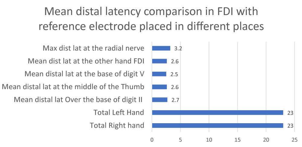
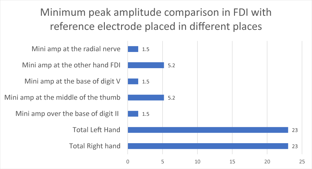
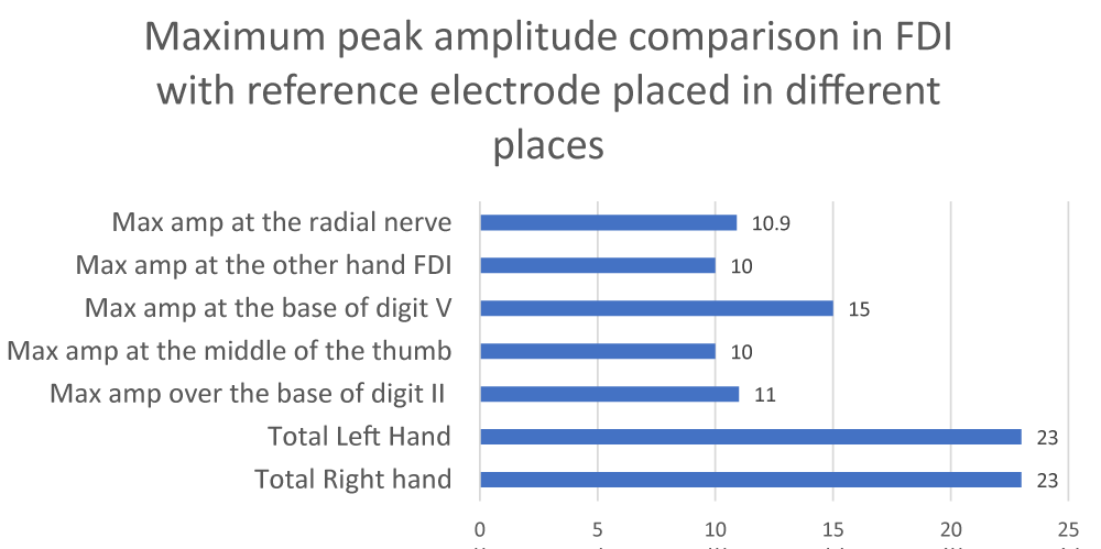
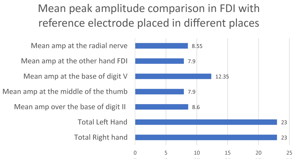
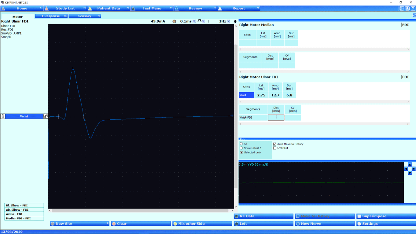
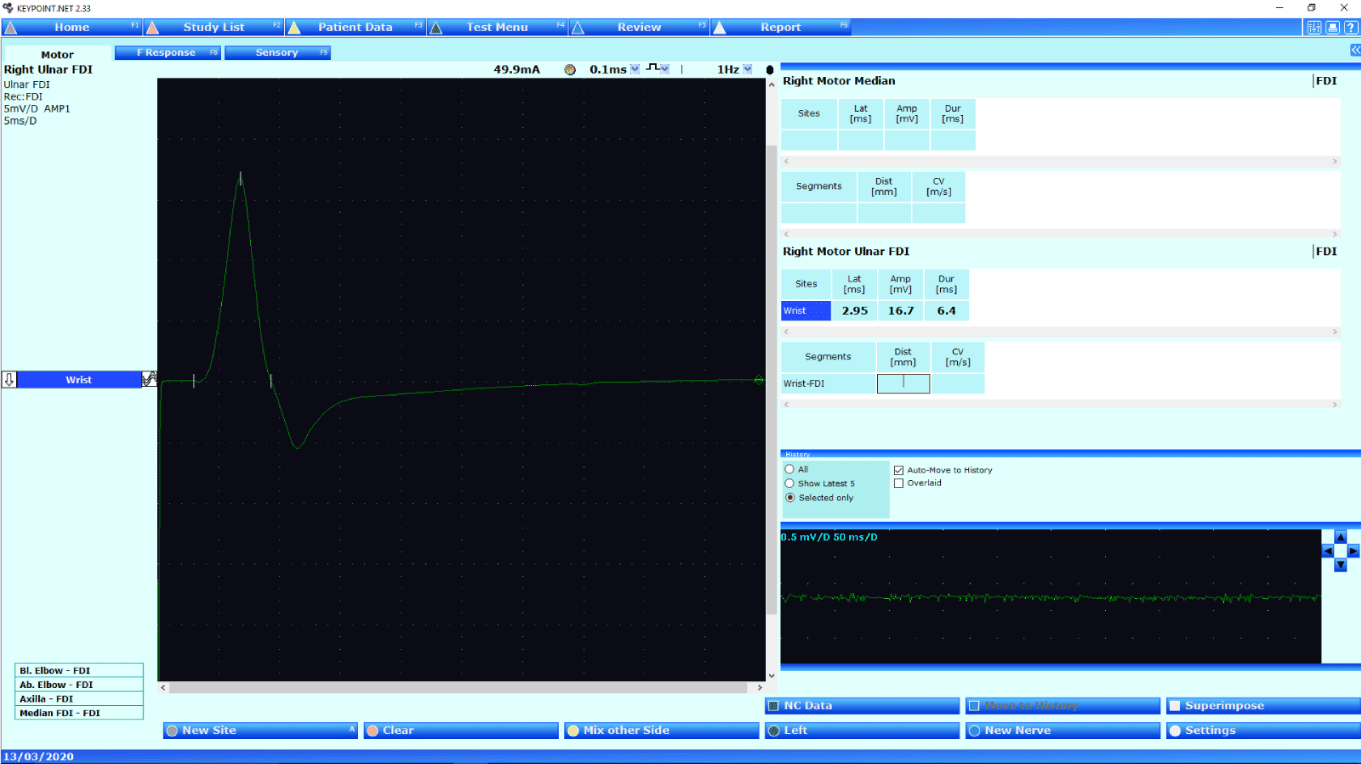
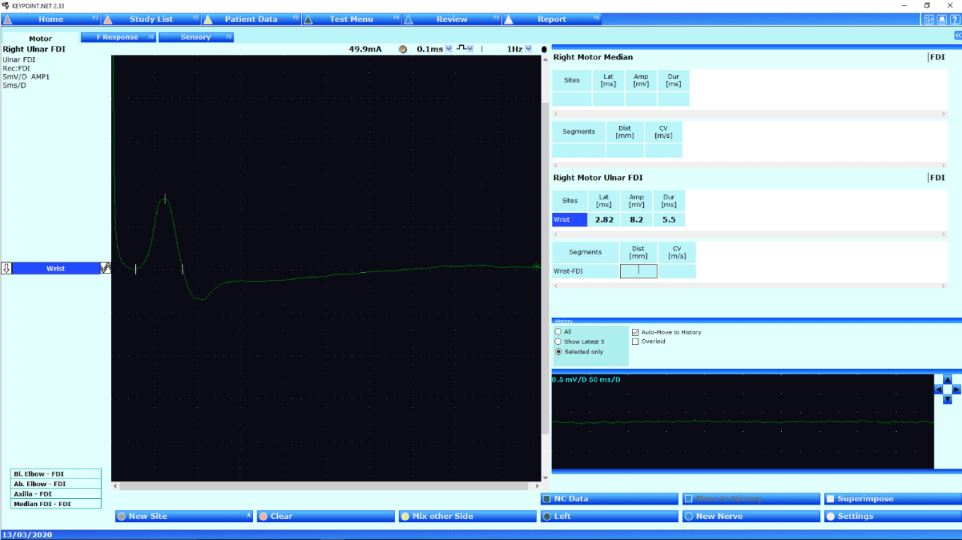
Patients aged 22-60 years, with a mean age of 19 years, were studied. Bilateral recordings were performed to ensure consistency in data collection (Graphs 1-6)(Pictures 1-9)(Tables 1-7).
The graph illustrates the baseline shift and amplitude difference.
Various published books and literature have recommended that the distance between the reference point and the active point in the stimulator may affect the recording amplitude. According to the literature, the ideal distance between the active and reference points in the stimulator should be 2 cm apart. In my experiments, I applied this principle to all 46 hands by placing the active and reference stimulating points at distances of 2 cm and 4.5 cm respectively, while positioning the active electrode on the FDI muscle and the reference electrode over the base of digit II, specifically the belly of the FDI muscles [16,17].
Results
Results indicate that all 46 hands had distal latency within normal limits, i.e., less than 4ms. All 46 hands exhibited normal amplitude, i.e., more than 5 mV. The comparison of amplitude data shows that the highest amplitude was recorded with the reference electrode at the base of digit V, corresponding to the tendon of the ADM muscles. The second-highest amplitude occurred when the reference electrode was placed at the base of digit II, corresponding to the tendon of the FDI muscles. The only difference between these positions is the baseline, where the base of digit II shows a positive deflection, while the base of digit V shows a clear distal latency baseline from which distal latency was calculated. Other reference electrode positions showed similar distal latency and amplitude, with slight elevated baselines for distal latency calculation. It was also observed that recording from FDI muscles at the belly with the reference electrode at the base of digit V showed a Martin-Gruber anomaly was observed 30% less frequently compared to recordings with the reference electrode at the base of digit II. The Martin-Gruber anomaly was not observed, when the elbow was bent at 900 and stimulating below the elbow, with the reference electrode at the base of digit II. This discrepancy often arises when clinical physiologists fail to bend the patient’s elbow during testing (CP) conduct studies without bending patients’ elbow at 900. The study conducted with the reference electrode at the base of digit V while recording from the FDI muscle indicates that if the elbow is not bent at 900, the Martin-Gruber phenomenon may still be detected if present. Results show no amplitude difference when the active electrode and the reference electrode of stimulation were placed at 2 cm and 4.5 cm while recording from FDI muscles with the reference electrode at the belly of FDI muscles [18,19].
Conclusion
Despite the limited sample size, the findings were consistent and clinically informative. The optimal electrode configuration for accurate CMAP recording from FDI muscles is by placing the reference electrode at the base of digit V, specifically at the tendon of the ADM muscle. These results confirm that the reference electrode influences CMAP readings and cannot be considered electrically inert. While conducting nerve conduction studies. The reference electrode contributes substantially to CMAP configuration during nerve conduction studies, and it also acts as an active electrode. Likewise, it is important to be consistent in using electrode placement when comparing studies and deciding on normal values. Recording from FDI muscles, by placing an active electrode and the reference electrode placed over the tendon of ADM muscles, offers improved clarity regarding the Martin-Gruber anomaly. The results indicate that Clinical physiologists may apply any commercially available stimulation devices on the market when recording FDI muscles.
Author’s contribution
The Author was responsible for collecting, analyzing, and interpreting data, as well as writing the manuscript.
Special thanks
I would like to thank my team who helped and supported me in different ways in completing this research. Special thanks to Dr. Bashir Kassam and Dr. Gareth Payne for their guidance in refining the manuscript.
- Joint ANS & BSCN audit for ulnar nerve entrapment. Available from: https://www.ansuk.org/asite/wp-content/uploads/2018/03/2014-ulnar.pdf
- Phongsamart G. Effect of reference electrode position on the compound muscle action potential (CMAP) onset latency. Muscle Nerve. 2002;25(6):816–21. Available from: https://doi.org/10.1002/mus.10119
- Optimal E2 (reference) electrode placement in fibular motor nerve conduction studies recording from the tibialis anterior muscle. Muscle Nerve. 2018. Available from: https://doi.org/10.1002/mus.26366
- Reference electrodes. In: Scholz F, editor. Electroanalytical methods. 2nd ed. Berlin: Springer; 2010; Chapter 15. Available from: http://ndl.ethernet.edu.et/bitstream/123456789/17131/1/Fritz%20Scholz.pdf
- Tankisi H, Burke D, Cui L, de Carvalho M, Kuwabara S, Nandedkar SD, et al. Standards of instrumentation of EMG. Clin Neurophysiol. 2020;131(1):243–58. Available from: https://doi.org/10.1016/j.clinph.2019.07.025
- Caress JB. Technical, physiological and anatomic considerations in nerve conduction studies. In: Blum AS, Rutkove SB, editors. The Clinical Neurophysiology Primer. Totowa (NJ): Humana Press; 2007. p. Chapter 13. Available from: http://dx.doi.org/10.1007/978-1-59745-271-7_13
- Kincaid JC, Brasher A, Markand ON. The influence of the reference electrode on CMAP configuration. Muscle Nerve. 1993;16:392–6. Available from: https://doi.org/10.1002/mus.880160408
- Brashear A, Kincaid JC. The influence of the reference electrode on CMAP configuration: leg nerve observations and an alternative site. Muscle Nerve. 1996;19:63–7. Available from: https://doi.org/10.1002/(sici)1097-4598(199601)19:1%3C63::aid-mus8%3E3.0.co;2-6
- Kimura J. Principles of nerve conduction studies. In: Kimura J, editor. Electrodiagnosis in diseases of nerve and muscle: principle and practice. 2nd ed. Philadelphia: FA Davis; 1983;33:83–4.
- Oh SJ. Nerve conduction techniques. In: Oh SJ, editor. Clinical electromyography: nerve conduction studies. 2nd ed. Baltimore: Williams & Wilkins. 1993;41–5.
- Weber RJ. Conduction and entrapment syndromes. In: Johnson EW, editor. Practical electromyography. Baltimore: Williams & Wilkins. 1988;111.
- Dumitru D, Amato AA, Zwarts MJ. Electrodiagnostic medicine. Philadelphia: Hanley & Belfus. 2002.
- Peckham PH, Knutson JS. Functional electrical stimulation for neuromuscular applications. Annu Rev Biomed Eng. 2005;7:327–60. Available from: https://doi.org/10.1146/annurev.bioeng.6.040803.140103
- Falco FJE, Hennessey WJ, Braddom RL, Goldberg G. Standardized nerve conduction studies in the upper limb of the healthy elderly. Am J Phys Med Rehabil. 1992;71:263–71. Available from: https://doi.org/10.1097/00002060-199210000-00003
- Eduardo E. The optimal recording electrode configuration for compound sensory action potentials. J Neurol Neurosurg Psychiatry. 1988;51:684–7. Available from: https://doi.org/10.1136/jnnp.51.5.684
- Nandedkar SD. Influence of reference electrode position on the compound muscle action potential. Clin Neurophysiol. 2020;131:160–6. Available from: https://doi.org/10.1016/j.clinph.2019.11.008
- Kimura J. Electrodiagnosis in diseases of nerve and muscle: principle and practice. 4th ed. Philadelphia: Oxford University Press. 1063.
- Brashear A, Kincaid JC. The influence of the reference electrode on CMAP configuration: leg nerve observations and an alternative site. Muscle Nerve. 1996;19:63–7. Available from: https://doi.org/10.1002/(sici)1097-4598(199601)19:1%3C63::aid-mus8%3E3.0.co;2-6
- Nandedkar SD. Contribution of reference electrode to the compound muscle action potential. Muscle Nerve. 2007;36:87–92. Available from: https://doi.org/10.1002/mus.20798
Article Alerts
Subscribe to our articles alerts and stay tuned.
 This work is licensed under a Creative Commons Attribution 4.0 International License.


 Save to Mendeley
