Journal of Neurology, Neurological Science and Disorders
Orbital Meningoencephalocele Due to Extraventricular Neurocytoma: Case Report
Andrea Cattalani1,2*, Vincenzo Grasso1, Matteo Vitali1, Gian Paolo Longo1, Narciso Mariani3 and Andrea Barbanera1
2Department of Clinical Surgical, Diagnostic and Pediatric Sciences, Neurosurgery, University of Pavia, Viale Golgi 19, 27100, Pavia, Italy
3Department of Services, Pathology Unit, Hospital SS. Antonio and Biagio and Cesare Arrigo, Via Venezia 16, 15121, Alexandria, Italy
Cite this as
Cattalani A, Grasso V, Vitali M, Longo GP, Mariani N, et al. (2016) Orbital Meningoencephalocele Due to Extraventricular Neurocytoma: Case Report. J Neurol Neurol Sci Disord 2(1): 007-009. DOI: 10.17352/jnnsd.000008Extraventricular Neurocytoma (EVN) is a rare primary tumor of Central Nervous System (CNS). To date, no cases have been reported in International Literature, about EVN associated to meningoencephalocele as manifestation of subacute increased intracranial pressure.
We report the first case of EVN that manifested with diplopia and ocular motor disorders due to intraorbital meningoencephalocele through a bone gap in the orbital roof.
Diagnosis of EVN is challenging and its differential diagnosis with more aggressive lesions is mandatory. Complete surgical removal does not need any adiuvant therapies and permits to control tumor growth and symptoms related to intracranial pressure.
Introduction
Extraventricular Neurocytoma (EVN) is a rare primary tumor of Central Nervous System (CNS) [1], -grade II according to World Health Organization (WHO) classification [2,3], that commonly manifests in young adults with partial seizures, headache or focal neurological deficits [4-6]. Conversely, Central Neurocytoma tipically presents with signs and symptoms of intracranial increased pressure and hydrocephalus [6]. To date, no cases have been reported in International Literature about EVN associated to bone erosion and parenchimal herniation as manifestations of subacute-chronic intracranial hypertension.
We report a case of extraventricular neurocytoma that manifested with diplopia and ocular motor disorders due to intraorbital increased pressure caused by meningoencephalocele through partial erosion of orbital roof.
Case Report
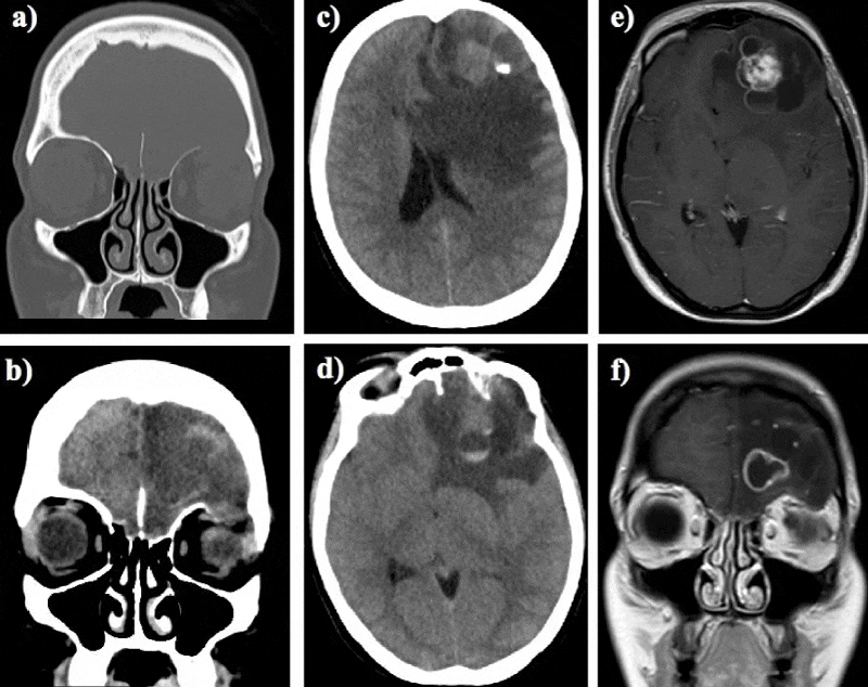
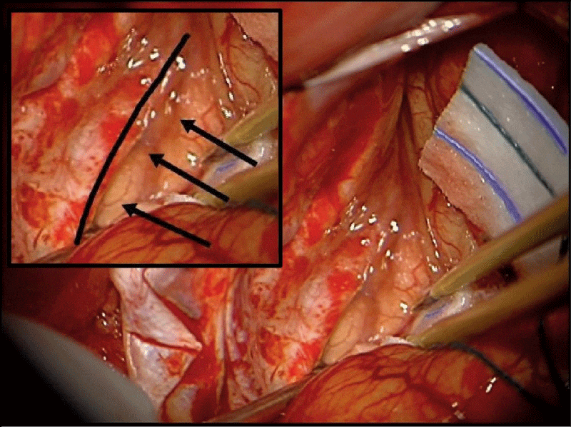
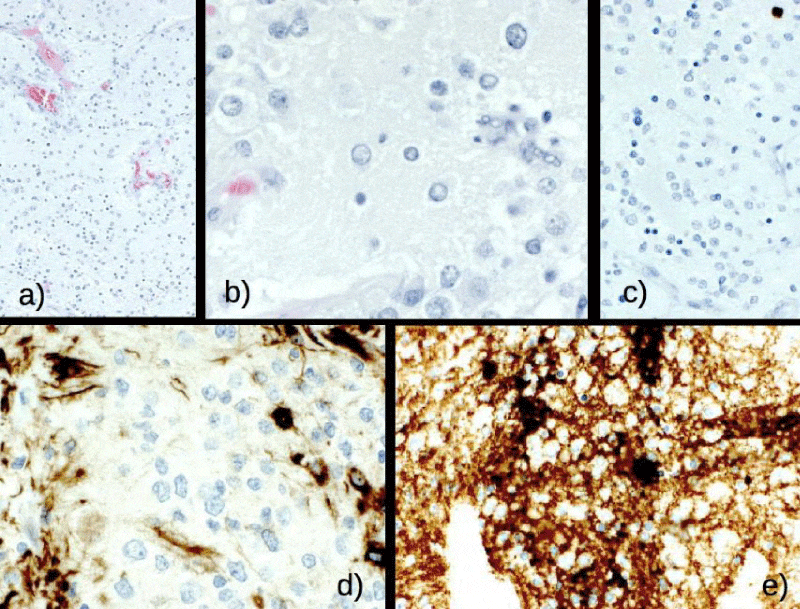
The patient, a 31-years old female, started to complain of mild headache and paroxysmal diplopia about one month before admission. Therefore, she underwent brain Magnetic Resonance Imaging (MRI) scan that showed the presence of a left frontal intraparenchimal lesion, with mixed solid and cystic components -with inhomogenous contrast-enhancement- and severe perilesional oedema. She was admitted to our Neurosurgical Department and neurological examination highlighted slight deficit of left estrinsic ocular motility and left exophthalmos; Computed Tomography (CT) brain scan also found signs of recent intra-tumoral hemorrhage, the presence of a small hyperdense calcification and bone-setting sequences revelead a small bone gap in the orbital roof (Figure 1). Patient underwent complete surgical removal of the lesion by left frontal craniotomy. Intraoperatively, a meningoencephalocele -dural and left frontal lobe herniation- was seen through a bone gap with smooth edges in the orbital roof, causing mass effect on intraorbital structures (Figure 2); the bone gap was repaired with two-layer heterologous dural patch and fibrin glue, with no direct bony reconstruction or lumbar drain. No intra or post-operative complications occurred. Serial post-operative CT and MRI scans with contrast showed complete excision of the mass. Patient referred complete recovery of pre-operative ocular disorders and progressive decrease of exophthalmos and headache. She was discharged home on 7th post-operative day. Hystological analysis confirmed diagnosis of Extraventricular Neurocytoma (WHO grade II) [2,3], with low proliferation index (Ki67 <1%), mitoses < 3 per 10 HPF. Immunohistochemical analysis found out clusts of cells with positivity for synaptophysin. No co-deletion 1p19q was found (Figure 3).
No further treatments were necessary. 1-month MRI follow up excluded neoplastic residuals or signs of recurrence.
Discussion
Extraventricular neurocytoma (EVN) is a rare intraparenchimal primary tumour of the Central Nervous System (CNS) -incidence about 0,13% [1], distinct hystological entity from the classic Central Neurocytoma; this latter one is typically located in the anterior portion of third and lateral ventricles, while EVN can be usually found in frontal and parietal lobes [7]. 2007 World Health Organization (WHO) classification first introduced EVN as a defined primary lesion of the CNS, included in the group of “neuronal and mixed neuronal-glial tumors” [3], further confirmed by 2016 WHO upgrade [2].
Clinical onset of EVN is typically characterized by partial seizures, headache, neurological focal deficits and less frequently by tumoral hemorrhage [4-6], distinguishing it from central neurocytoma that commonly causes symptoms of increased intracranial pressure or hydrocephalous. This nonspecific clinical manifestation does not allow to make any definitive diagnostic hypothesis. Furthermore, also EVN radiological features are not pathognomonic. In fact it may be seen as a well-circumcribed round or multilobular mass, with frequent cystic degeneration areas, mural nodules and calcifications; Magnetic Resonance Imaging (MRI) T1-weighted sequences typically show isointense or slighlty hyperintense signal with heterogeneous contrast-enhancement. Common differential diagnosis is with oligodendroglioma, ependymoma and dysembryoplastic neuroepithelial tumors [1].
Histologically EVN is made up by a composition of monotonous neoplastic cells with round, regular nuclei embedded in a matrix of fine neuronal processes, known as neuropils -finer than glial processes [8]. Atypical EVN presents a proliferation index (MIB-1, Ki-67) of >2%, atypical features -such as necrosis, neoangiogenesis or mitoses > 3 per 10 HPF [9], and more aggressive neurobiological behavior in respect of central neurocytoma. Immunohistochemical analysis presents high synaptophysin positivity and in some cases there is 1p19q codeletion, tipically carried by oligodendrogliomas.
Complete surgical resection is currently the “gold standard” treatment [10], in cases of sub-total removal, radiotherapy may be a valid alternative to control tumor growth [11]. To date, no supported data can be found about radiosurgery rate of activity on EVNs.
Meningoencephalocele (MEC) is the herniation of brain tissue, covered by meninges, via openings of the skull base [12], it can be caused by surgery, traumas, neoplasia, infections or may occur spontaneously and sometimes it can be associated to cerebrospinal fluid fistulas. Exact mechanism of MECs in patients with intracranial tumors is debatable, although two different ways can be involved: the “direct” one with dural and bone erosion directly by tumor and the “indirect” one, in which it is possible that subacute-chronic effect of local intracranial hypertension and pulsations on fragile areas of skull base may cause gradual thinning and loss of integrity of the bone [12,13].
To date, no cases of MEC caused by EVN-related intracranial hypertension have been reported in International Literature.
In the present patient, EVN caused severe peritumoral oedema and progressive increase of intracranial pressure, determining herniation of left frontal lobe and dura mater through a bone gap in the omolateral orbital roof; although it is not possible to demonstrate any causative relation between intracranial hypertension and erosion of orbital roof, the bone gap was homolateral to the EVN and had smooth edges: these elements and the integrity of dura mater let us to suspect that intracranial hypertension related to EVN may have caused not only the herniation through the bone gap, but also the formation of the same gap. Complete surgical excision of the lesion permitted us to solve any patient symptom -especially those linked to intraorbital mass effects and intracranial hypertension- and do not need any further treatment.
Conclusion
This is the first case of EVN presenting with signs of intracranial hypertension causing herniation of cerebral parenchima through a bone acquired gap. Diagnosis of EVN is challenging and differential diagnosis with more aggressive lesions is mandatory, because complete surgical removal does not need any adiuvant therapies and permits to control, as in our patient, mass effect on intra and extra-cranial structures.
Informed Consent
The patient has consented to submission of this case report to the journal.
- Liu K, Wen G, Lv XF, Deng YJ, Deng YJ, et al. (2013) MR imaging of cerebral extraventricular neurocytoma: A report of 9 cases. AJNR Am J Neuroradiol 34: 541-546. Link: https://goo.gl/0WwQel
- Louis DN, Perry A, Reifenberger G, von Deimling A, Figarella-Branger D, et al. (2016) The 2016 World Health Organization Classification of Tumors of the Central Nervous System: a summary. Acta Neuropathol. 131: 803-820. Link: https://goo.gl/ncaiPr
- Sharma MC, Deb P, Sharma S and Sarkar C (2006) Neurocytoma: A comprehensive review. Neurosurg Rev 29: 270-285 Link: https://goo.gl/vS3mNo
- Ji YC, Hu JX, Li Y, Yan PX, Zuo HC (2016) Extraventricular neurocytoma in the left temporal lobe: A case report and review of the literature. Oncol Lett 11: 3579-3582. Link: https://goo.gl/mvMvG0
- Messina R, Cefalo MG, Secco DE, Cappelletti S, Rebessi E, et al. (2014) Behavioral disorders as unusual presentation of pediatric extraventricular neurocytoma: report on two cases and review of the literature. BMC Neurol 19: 242. Link: https://goo.gl/z6suTH
- Taylor CL, Cohen ML, Cohen AR (1998) Neurocytoma presenting with intraparenchymal cerebral hemorrhage. Pediatr Neurosurg 29: 92-95. Link: https://goo.gl/2AqmfD
- Hassoun J, Gambarelli D, Grisoli F, Pellet W, Salamon G, et al. (1982) Central neurocytoma. An electron-microscopic study of two cases. Acta Neuropathol 56: 151-156. Link: https://goo.gl/vTgkFB
- Brat DJ, Scheithauer BW, Eberhart CG, Burger PC (2001) Extraventricular neurocytomas: Pathologic features and clinical outcome. Am J Surg Pathol 25: 1252-1260. Link: https://goo.gl/hgM3tN
- Rades D, Fehlauer F, Schild SE (2004) Treatment of atypical neurocytomas. Cancer 100: 814-817. Link: https://goo.gl/8VBMaZ
- Rades D, Schild SE, Fehlauer F (2004) Defining the best available treatment for neurocytomas in children. Cancer 101: 2629-2632. Link: https://goo.gl/uFbgqH
- Patil AS, Menon G, Easwer HV, Nair S (2014) Extraventricular neurocytoma, a comprehensive review. Acta Neurochir (Wien) 156: 349-354. Link: https://goo.gl/chWpGv
- Papanikolaou V, Bibas A, Ferekidis E, Anagnostopoulou S, Xenellis J (2007) Idiopathic temporal bone encephalocele. Skull Base. 17: 311-316. Link: https://goo.gl/mL0yLr
- Ommaya AK, Di Chiro G, Baldwin M, Pennybacker JB (1968) Non-traumatic cerebrospinal fluid rhinorrhoea. J Neurol Neurosurg Psychiatry 31: 214-225. Link: https://goo.gl/ZBuL4v
Article Alerts
Subscribe to our articles alerts and stay tuned.
 This work is licensed under a Creative Commons Attribution 4.0 International License.


 Save to Mendeley
