Journal of Neurology, Neurological Science and Disorders
Prevalence of autoimmune encephalitis in patients with a first episode psychosis
Mataró Hospital, Maresme Health Consortium, Mataró, Spain
Author and article information
Cite this as
Remón MC, Ruiz VC, Batiste DM, Buscà NG, Serven EG, et al. (2020) Prevalence of autoimmune encephalitis in patients with a first episode psychosis. J Neurol Neurol Sci Disord. 2020; 6(1): 10-13. Available from: 10.17352/jnnsd.000038
Copyright License
© 2020 Remón MC, et al. This is an open-access article distributed under the terms of the Creative Commons Attribution License, which permits unrestricted use, distribution, and reproduction in any medium, provided the original author and source are credited.Autoimmune encephalitis are a group of inflammatory diseases of the central nervous system whose clinical presentation uses to be characterized by neuropsychiatric disorders. Different autoantibodies against neuronal cell-surface proteins and synaptic receptors have been described as causing autoimmune encephalitis. Of these, anti-N-methyl-Daspartate receptor (anti-NMDAR) encephalitis is the most frequent, with an estimated incidence of 1.5 per million inhabitants/year. Other antibodies have also been linked to autoimmune encephalitis,
Autoimmune encephalitis are a group of inflammatory diseases of the central nervous system whose clinical presentation uses to be characterized by neuropsychiatric disorders. Different autoantibodies against neuronal cell-surface proteins and synaptic receptors have been described as causing autoimmune encephalitis. Of these, anti-N-methyl-Daspartate receptor (anti-NMDAR) encephalitis is the most frequent, with an estimated incidence of 1.5 per million inhabitants/year. Other antibodies have also been linked to autoimmune encephalitis, such as intraneuronal antibodies (clearly associated with neoplastic diseases), antithyroid antibodies, and those related to systemic diseases such as lupus. The focus of this study does not include these antibodies since there is often additional symptomatology that facilitates diagnosis and specific analytical screening is conducted when there is a clinical suspicion.
Symptoms often consist in cognitive-behavioral, mood, and psychotic disorders, especially in limbic and anti-NMDAR encephalitis. This means that, in many cases, patients with encephalitis may be initially treated in psychiatric wards for probable first-episode psychosis. If not treated properly, the course of illness is often unfavourable and admission to intensive care units may be necessary due to altered consciousness, status epilepticus or hemodynamic instability derived from severe dysautonomia. However, there have been cases of atypical (mild or attenuated) encephalitis characterized primarily by symptoms of psychosis and sometimes accompanied by mild neurological symptoms and/or dysautonomia. This type of encephalitis uses to take a more benign course than “classic” autoimmune encephalitis. While it is characteristically refractory to antipsychotic medication, immunotherapy may be beneficial in such cases, which makes its early diagnosis of increasing interest.
The diagnostic approach to autoimmune encephalitis follows the criteria of Graus, et al. [1], to identify clinically recognizable syndromes and, thus, start immunotherapy early. Brain MRI, CSF analysis and EEG studies are used as complementary tests, among others. Typical brain MRI shows hyperintense lesions on T2-weighted FLAIR sequences restricted to both medial temporal lobes (in limbic encephalitis) or multifocal corticosubcortical lesions suggesting demyelination or inflammation. CSF analysis may show mild pleocytosis and/or high CSF protein concentrations. EEG studies often present both nonspecific abnormalities (from slowing to focal epileptiform activity) and specific abnormalities (in the case of extreme delta brush in anti-NMDAR encephalitis). That said, probable autoimmune encephalitis may not be ruled out merely due to normal results on these tests. A combination of immunohistochemistry and cell-based assays must also be employed to detect antibodies in the blood and CSF. The presence of antibodies is essential to determine the etiology and, in turn, the prognosis, comorbidities and associated tumors. It should be noted that the presence of negative antibodies in the right clinical context does not preclude a diagnosis of autoimmune encephalitis.
While the true prevalence of autoimmune encephalitis is currently unknown, it is considered the third most common cause of encephalitis, following infections (especially viruses) and acute disseminated encephalomyelitis [2]. Over the past years, it has had an incidence similar to that of other types of viral encephalitis [3].
The literature describes patients case series with autoimmune encephalitis and questions whether it might be underdiagnosed in our setting [4]. Moreover, there is some recognition of the existence of autoimmune psychosis [5] in the field of psychiatry after finding that a variable proportion of patients with symptoms of psychosis were AntiNMDA R positive. It must be noted, however, that diagnosis is confirmed via positive results for antibodies in CSF obtained with appropriate techniques, and a correlation with Anti NMDA R positivitiy in serum/CSF is not always found [6].
In light of this, we believe that proper CSF analysis in suspected patients is crucial. In this study, we include CSF analysis as an essential test to diagnose autoimmune encephalitis.
Aim of the study
This study aims to determine the prevalence of autoimmune encephalitis in patients with first-episode psychosis by means of a joint care protocol employed by psychiatrists and neurologists and to assess the advantages of implementing it in clinical practice.
Material and methods
The general hospital this study was conducted at is a center of reference in psychiatric care and serves 400,000 inhabitants. Neurologists and psychiatrists worked together to develop a protocol of early, joint and systematic care for patients experiencing a first episode of psychosis.
Following the implementation of the protocol on 1 May 2018, the patients evaluated were consecutively included in an observational study. Clinical and paraclinical data were collected prospectively from immunological blood testing, CSF analysis, MRI and EEG.
Patints at our center were included in the study consecutively, as they arrive to emergency psiquiatric ward. The only inclusion criterion was that they had experienced a first episode of psychosis within the last six months. Exclusion criteria were substance-induced psychosis, prior psychotic disorders, neurodegenerative diseases, and difficulty understanding and/or inability to complete follow-up. The whole project consisted of two studies:
-The first was a cross-sectional studie that involved the evaluation of patients through neurological examination, neuropsychological assessment, serum analysis, brain MRI, EEG and lumbar puncture. Blood and CSF samples were sent to the neuroimmunology unit at Hospital Clínic de Barcelona for the detection of antibodies to neuronal cell-surface antigens using immunohistochemistry and cell-based assays.
-The second was a prospective studie involving clinical and functional assessment at follow-up at two, four, six and 12 months.
All patients were informed of the option to voluntarily participate in the study and gave their informed consent if they decided to join. The study was approved by the Ethics Committee at our center (Consorci Sanitari del Maresme).
Results
Over 18 months (May 2018 - November 2019), 75 patients (37 female, 38 male) with a mean age of 35 (15 - 75) were included in the study. Seven patients were excluded: five due to refusal to participate, one to intellectual disability and one to substance-induced psychosis.
-Brain imaging results: Brain MRI was performed on 64 of the 75 patients, with 25 presenting nonspecific findings. In one patient, non-Gadolinium enhanced hyperintense corticosubcortical lesions were observed in the orbitofrontal region and temporal lobe, suggesting an encephalitic process. Despite the detection of negative antibodies, the patient presented an unfavourable course of illness associated with symptoms of dysautonomia and failure to respond to antipsychotic treatment, which initially led to suspicions of probable autoimmune encephalitis with negative antibodies based on the criteria of Graus, et al. [1]. Immunotherapy was therefore initiated. Ultimately, however, the same MRI abnormalities were detected in the patient’s sister, who presented abnormal behavior. which leaded to the diagnosis suspicion of an hereditary metabolic disease.
-EEG results: EEG was performed on 59 patients. Nonspecific diffuse slowing was found in two patients and bifrontal epileptiform activity was detected in one patient.
-CSF analysis results: Lumbar puncture was performed on all 75 patients. The results of the biochemical analysis showed <5 cells in all patients and high CSF protein concentrations in one patient, who was eventually diagnosed with cervical spinal tuberculosis and myelopathy. The detection of antibodies using immunohistochemistry and cell-based assays obtained negative results in all patients. However, in one case, a commercial kit was used to detect antibodies in a patient at another center and yielded positive results, which initially led to a false positive. Once transferred to our center, the patient showed improvement without receiving immunotherapy. Repetition of the tests via immunohistochemistry and cell-based assays at the Hospital Clínic immunology lab did not produce positive results.
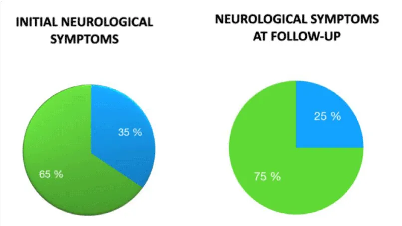
-Neurological examination results: At the initial neurological examination, 35% of patients presented neurological symptoms (some of them more than one), the most frequent being insomnia and abnormal movements, especially tremor. In follow-up examinations at four months (available in 48 pacients), 25% presented neurological symptoms, with the most frequent being headache and movement disorders. See table.
Discussion
This study analyzed data from 75 patients experiencing first-episode psychosis who consecutively came to our center over 18 months, in an attempt to estimate the prevalence of autoimmune encephalitis that might be underdiagnosed in patients normally examined in psychiatric wards.
The study was conducted under optimal conditions for the detection of antibodies, in serum and CSF, also included exhaustive follow-up of participants, and it did not identify any patients with autoimmune encephalitis. Although the results are negative, we believe this finding is relevant since it supports existing data on the infrequency of this type of pathology and the importance of presenting compatible neurological symptoms and course of illness. The history of the disease and correct interpretation of complementary tests, such as MRI, remain vitally important to preventing false positives such as those described above. We also believe that the detection of antibodies should be conducted by experienced labs using immunohistochemistry and cell-based assays.
The results of this study corroborate the idea that autoimmune encephalitis characterized primarily by psychotic symptoms is very infrequent and complex to diagnose. However, given the response of this entity to immunotherapy and/or excision of the associated tumor, approaching it correctly has become increasingly important and several articles have recently been published on the topic [7]. Pollack, et al. [5], for example, put forth the concept of autoimmune psychosis, as well as a series of warning signs, and diagnostic criteria for possible, probable, and definite autoimmune encephalitis. It’s also worth mentioning a study by Servén et al. [8], in which the similarities between anti-NMDAR encephalitis and cycloid psychosis are presented in three cases of females with C-cluster personality traits and first-episode psychotic symptoms triggered by a stressor. All three patients met the diagnostic criteria for cycloid psychosis but presented adverse effects and poor evolution when treated with antipsychotics. This led to suspicions and, eventually, confirmation of anti-NMDAR encephalitis related to ovarian teratoma.
Another study, by Kayser, et al. [9], described the findings of paraclinical tests in patients with anti-NMDAR encephalitis, demonstrating their non-specific nature. This reveals the importance of conducting proper CSF analysis, as in this study. More recently, Dalmau, et al. [10], published a new update on anti-NMDAR encephalitis aimed at psychiatrists and neurologists. Under the acronym SEARCH For NMDAR-A, they proposed new diagnostic keys for anti-NMDAR encephalitis in patients with first-episode psychosis.
Conclusions
Autoimmune encephalitis is an infrequent disease. We have not detected any cases of it to date, which supports its low incidence. The use of reliable lab techniques to prevent false positives/negatives and clinical follow-up of patients, especially in the event of unclear diagnosis, is essential. Warning signs associated with psychotic disorders that suggest autoimmune encephalitis must be detected in order to suspect diagnosis. Moreover, joint work between neurologists and psychiatrists is decisive for optimal management of the disease. More studies should be conducted to deepen existing knowledge on this pathology, the current approach to it and the different ways it presents to optimize treatment and prognosis. In this regard, there are several studies currently underway, such the one conducting at the moment at Hospital Clinic (Barcelona) on the incidence of autoimmune encephalitis in patients with psychotic symptoms, which will offer results soon.
Dr. Dalmau, Dr. Graus and Dr. Guasp (Hospital Clínic, Barcelona).
- Graus F, Titulaer MJ, Balu R, Benseler S, Bien CG, et al. (2016) A clinical approach to diagnosis of autoimmune encephalitis. Lancet Neurol 15: 391-404. Link: https://bit.ly/2D6052t
- Granerod J, Ambrose HE, Davies NW, Clewley JP, Walsh AL, et al. (2010) Causes of encephalitis and differ- ences in their clinical presentations in England: a multicentre, population-based prospective study. Lancet Infect Dis 10: 835-844. Link: https://bit.ly/31H3EXg
- Dalmau J, Graus F (2018) Antibody-mediated encephalitis. N Engl J Med 378: 840-851. Link: https://bit.ly/31FOWQc
- Prüss H, Dalmau J, Harms L, Höltje M, Ahnert-Hilger G, et al. (2010) Retrospective analysis of NMDA receptor antibodies in encephalitis of unknown origin. Neurology 75: 1735-1739. Link: https://bit.ly/2NRwDiO
- Pollak TA, Lennox BR, Müller S, Benros ME, Prüss H, et al. (2019) Autoimmune psychosis: an international consensus on an approach to the diagnosis and management of psychosis of suspected autoimmune origin. Lancet Psychiatry 7: 93-180. Link: https://bit.ly/2YVEfad
- Scott JG, Gillis D, Ryan AE, Hargovan H, Gundarpi N, et al. (2018) The prevalence and treatment outcomes of antineuronal antibody- positive patients admitted with first episode of psychosis. BJPsych Open 4: 69–74. Link: https://bit.ly/2VEST3y
- Guanyabens-Buscà N, Casado- Ruiz V, Giné-Serven E, Palomeras E, Muriana D, et al. (2019) Encefalitis antirreceptor de NMDA. Diagnóstico y tratamiento precoz en pacientes con sintomatología psicótica aguda-subaguda. Rev Neurol 68: 18-22. Link: https://bit.ly/2YXv3Cu
- Giné Servén E, Quintana EB, Buscà NG, Ruiz VC, Rivas CT, et al. (2019) Considerations of psychotic symptomatology in anti-NMDA encephalitis: Similarity to cycloid psychosis. Clin Case Rep 7: 2456-2461. Link: https://bit.ly/2C3rUrs
- Kayser MS, Titulaer MJ, Gresa-Arribas N, Dalmau J (2013) Frequency and characteristics of isolated psychiatric episodes in anti-N-methyl-d-aspartate receptor encephalitis. JAMA Neurol 70: 1133–1139. Link: https://bit.ly/3f8JXvn
- Dalmau J, Armangué T, Planagumà J, Radosevic M, Mannara F, et al. (2019) An update on anti-NMDA receptor encephalitis for neurologists and psychiatrists: mechanisms and models. Lancet Neurol 18: 1045-1057. Link: https://bit.ly/3eRiApg



 Save to Mendeley
Article Alerts
Subscribe to our articles alerts and stay tuned.
 This work is licensed under a Creative Commons Attribution 4.0 International License.