Archives of Depression and Anxiety
Crosstalk of Gastrointestinal Pathogen Infection, Food Allergy, and Neuroinflammation in Alzheimer’s Disease
1Molecular Virology and Microbiology, Baylor College of Medicine, One Baylor Plaza, Houston, T 77030, USA
2Department of Health & Biomedical Sciences, College of Health Professions, University of Texas Rio Grande Valley. One West University Blvd. Brownsville, TX 78520-4933, USA
Author and article information
Cite this as
Su BB, et al. Crosstalk of Gastrointestinal Pathogen Infection, Food Allergy, and Neuroinflammation in Alzheimer’s Disease. Arch Depress Anxiety. 2026; 12(1): 1-9. Available from: 10.17352/2455-5460.000105
Copyright License
© 2026 Su BB, et al. This is an open-access article distributed under the terms of the Creative Commons Attribution License, which permits unrestricted use, distribution, and reproduction in any medium, provided the original author and source are credited.Alzheimer’s Disease (AD), the most common cause of dementia worldwide, is underrecognised as a systemic disorder that extends beyond the central nervous system. Emerging evidence highlights the critical role of the Gut–Brain Axis (GBA), a bidirectional communication network linking intestinal microbes, immune responses, and neural pathways. Perturbations in this axis, such as enteric microbial dysbiosis and food allergy, may contribute to immune dysregulation, gut microbial alterations, and intestinal barrier dysfunction, which are hypothesised to influence β-amyloid aggregation, tau pathology, and ultimately cognitive decline. These insights underscore the importance of intestinal homeostasis in modulating susceptibility to neurodegeneration. Several factors contribute to the pathogenesis of AD; however, this review focuses on food allergies and Gastrointestinal Pathogens (GIPs). Despite decades of research, there is currently no curative treatment; existing pharmacological therapies provide only modest symptomatic relief and slow disease progression. Consequently, clinical interest has grown in complementary and integrative approaches, particularly traditional herbal medicines and acupuncture, which have long histories of use in maintaining systemic balance and preventing AD progression. Preclinical and clinical studies suggest that such interventions may regulate gut microbiota composition, enhance barrier integrity, suppress pro-inflammatory signalling, and confer direct neuroprotective effects. In this review, we discuss the therapeutic potential of traditional medicine-based interventions as adjunctive strategies for AD management, with emphasis on their ability to target the GBA, modulate immune responses, and improve neuroprotection. These perspectives may provide insight for future preventive and therapeutic frameworks for AD.
AD: Alzheimer’s Disease; GBA: Gut-Brain Axis: TLR: Toll-Like Receptor: IL: Interleukin; EED: Environmental Enteric Dysfunction; Aβ: Amyloid-β; BBB: Blood–Brain Barrier; FPIES: Protein-Induced Enterocolitis Syndrome; ILC2s: Type 2 Innate Lymphoid Cells; CSF: Cerebrospinal Fluid; EPEC: Entero Pathogenic E. coli; PI-IBS: Post-Infectious Irritable Bowel Syndrome; SCFAs: Short-Chain Fatty Acids; Treg: Regulatory T Cell; CNS: Central Nervous System; EGIDs: Eosinophilic Gastrointestinal Disorders; GP: Gut Pathogen; GIP: Gastrointestinal Pathogen; OBA: Organ-Brain Axis
Introduction
Alzheimer’s Disease (AD) is the most common cause of dementia worldwide, affecting more than 55 million people as of 2023, and its prevalence is characterised by progressive cognitive decline and behavioural impairment. AD has long been understood primarily as a Central Nervous System (CNS) disorder marked by extracellular amyloid-b (Ab) deposition, intracellular tau hyperphosphorylation, synaptic dysfunction, and neuronal death. Growing evidence implicates systemic factors—particularly immune dysfunction and gut disturbances in shaping AD risk and progression [1-3]. This review highlights the potential indirect roles of food allergy and GIP infection in the development of AD, distinct from established genetic and environmental influences.
Several FDA-approved drugs now target amyloid plaques in Alzheimer’s disease, including lecanemab (Leqembi) and donanemab (Kisunla). These therapies are indicated for patients in the early stages of Alzheimer’s and are designed to slow disease progression by promoting amyloid clearance from the brain. However, their use is limited by significant safety concerns. The most notable adverse effect is amyloid-related imaging abnormalities (ARIA), which may present as oedema (swelling) or microhemorrhages (bleeding). Additional side effects include headaches, dizziness, and infusion-related reactions. Given these challenges, there is an urgent need to develop safer and more effective strategies for the prevention and treatment of Alzheimer’s disease [4-6].
Current findings
Intestinal dysfuncionin AD
One of the key intestinal abnormalities in AD is gut barrier impairment. Both human and animal studies reveal increased intestinal permeability (“leaky gut”) in [7]. The Zou group found reduced expression of tight junction proteins (occludin, claudin-1, and ZO-1) in AD mouse models, which allowed bacterial products like Lipopolysaccharide (LPS) to enter systemic circulation [8]. Elevated LPS levels have been detected in postmortem AD brains and colocalise with Aβ plaques, suggesting translocation from the gut contributes to neuroinflammation [9]. Compromised gut integrity facilitates chronic systemic inflammation, which accelerates neuronal injury and cognitive decline.
Microbiota dysbiosis of AD
AD patients frequently exhibit altered gut microbiota composition. Metagenomic studies show reduced abundance of beneficial taxa such as Bifidobacterium and Faecalibacterium prausnitzii, alongside enrichment of proinflammatory species including Escherichia/Shigella [10]. These shifts reduce production of short-chain fatty acids (SCFAs) like butyrate, which normally maintain epithelial barrier integrity and support regulatory T cell (Treg) differentiation. Butyrate deficiency has been associated with increased neuroinflammation and accelerated Ab pathology [11]. Dysbiosis also alters bile acid metabolism, producing neurotoxic secondary bile acids implicated in cognitive decline.
Immune dysregulation of AD
Intestinal dysfunction in AD includes dysregulated mucosal immunity. Microbial metabolites and antigens activate gut immune cells, promoting a Th1/Th17-skewed response. This results in systemic release of IL-6, TNF-α, and IL-1β, which cross the blood–brain barrier (BBB) and exacerbate neuroinflammation; this latter can also be triggered by IL-8 and IL-18 [12,13]. Evidence also suggests that intestine -derived immune cells can migrate to the CNS. In APP/PS1 mice, activated intestinal T cells infiltrated the brain and worsened amyloid pathology [14]. Thus, intestinal immune dysregulation links peripheral dysfunction with central AD pathology.
In summary, intestinal dysfunction plays a critical role in AD pathogenesis by disrupting the Gut–Brain Axis (GBA), impairing barrier integrity, altering microbial composition, and driving immune dysregulation, which collectively promote neuroinflammation and accelerate the accumulation of amyloid and tau pathology.
Gastrointestinal pathogens
Infection may contribute to alzheimer’s disease pathogenesis through intestinal dysfunction
GIP infections, caused by bacteria, viruses, and parasites, remain a leading cause of gastrointestinal morbidity worldwide. Common pathogens include Escherichia coli, Salmonella, Shigella, Clostridium diffcile, rotavirus, norovirus, and protozoa such as Giardia lamblia. While many infections are acute and self-limiting, they often trigger persistent alterations in intestinal physiology. These infections may cause intestinal dysfunction, defined as impaired barrier integrity, altered immune signalling, intestinal dysbiosis, and long-term sequelae such as post-infectious irritable bowel syndrome (PI-IBS) or malabsorption, and Environmental Enteric Dysfunction (EED) in adults, as a result indirectly contributing to AD pathogenesis through intestinal, BBB, and GBA dysfunction.
Enteric pathogens lead to barrier dysfunction
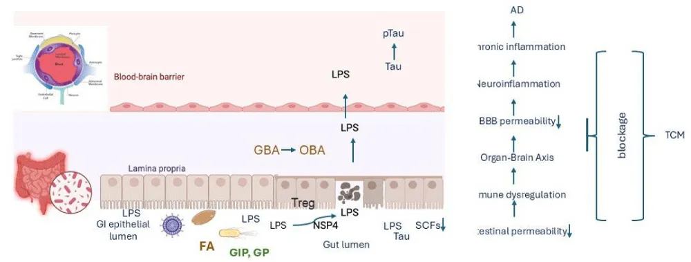
One major effect of enteric pathogens is damage to the epithelial barrier. Pathogens deploy toxins and virulence factors that disrupt tight junction proteins, increasing permeability (“leaky gut”). For example, Enteropathogenic E. coli (EPEC) injects effector proteins via a type III secretion system, leading to pedestal formation, epithelial injury, and barrier leakage (Martinez-Argudo et al., 2010). Similarly, C. difficile toxins A and B cause cytoskeletal disruption and epithelial apoptosis, resulting in codiarrhoea [15] and non-structured protein (NSP4) [16]. Barrier disruption permits translocation of bacterial products such as LPS into the lamina propria and circulation. This amplifies immune activation and perpetuates inflammation, contributing to chronic dysfunction beyond acute infection.
Enteric pathogens trigger immune dysregulation
Enteric infections provoke strong innate and adaptive immune responses. Pattern recognition receptors (PRRs) such as Toll-like receptors (TLRs) detect microbial products, activating nuclear factor kappa-light-chain-enhancer of activated B cells (NF-κB) and inflammasome pathways. This induces proinflammatory cytokines (IL-1β, IL-8, IL-18, TNF-α, IL-6) that recruit neutrophils and macrophages to infection sites [17,18] and may contribute to AD pathogenesis, as elevated levels of several of these cytokines have been detected in the serum and cerebrospinal fluid (CSF) of AD patients [12,13,19].
Enteric pathogens disrupt healthy intestinal microbiota
Also disturb the gut microbiota, resulting in dysbiosis. Antibiotic-associated C. difficile infection exemplifies how pathogen overgrowth following microbiome disruption causes recurrent colitis. Viral infections such as rotavirus also induce long-term shifts in microbial ecology, reducing beneficial commensals and enabling pathogen persistence [20] and also exacerbate EED in low-resource settings. Repeated childhood infections contribute to chronic villous blunting, impaired absorption, stunted growth, and poor oral vaccine responses [21]. Microbiota perturbation impairs colonisation resistance, facilitating recurrent infections and chronic inflammation. Furthermore, reduced production of SCFAs, such as butyrate, weakens epithelial barrier function and regulatory immune responses, reinforcing dysfunction.
In summary, enteric pathogens disrupt gut–brain axis homeostasis by damaging the intestinal barrier, inducing immune dysregulation, and reducing microbial diversity. These alterations stimulate the release of pro-inflammatory cytokines, facilitate their translocation across the BBB, and exacerbate neuroinflammation, thereby contributing to Alzheimer’s disease pathogenesis through systemic–central immune crosstalk.
Food allergy may contribute to alzheimer’s disease pathogenesis through intestinal dysfunction
Food allergy promotes asthma, atopic dermatitis, and immune-mediated adverse reactions to dietary antigens, affecting approximately 6–8% of children and up to 4% of adults worldwide. It can be life-threatening in anaphylaxis. The intestinal tract, as the primary site of exposure to food antigens, plays a central role in both the initiation and manifestation of allergic responses. Atopic diseases—including asthma, allergic rhinitis, and atopic dermatitis—are significantly and dose-dependently associated with increased dementia risk [22] and food allergy was also found to increase phosphorylation of tau protein in the brain [23].
Food allergy has recently been recognised as a driver of intestinal dysfunction, characterised by barrier disruption, altered microbial ecology, and dysregulated immune responses.
Food allergy may promote intestinal dysfunction
The intestinal epithelium forms a physical and biochemical barrier that regulates antigen passage from the lumen to systemic circulation. In food allergy, epithelial barrier integrity is compromised. Studies demonstrate increased intestinal permeability (“leaky gut”) in allergic individuals, attributed to disrupted tight junction proteins such as occludin and claudins [24,25]. This impaired barrier allows excessive penetration of food antigens and microbial products, perpetuating allergic sensitisation and systemic inflammation.
Additionally, epithelial cells in allergic intestines often secrete higher levels of thymic stromal lymphopoietin (TSLP) and IL-33, which activate dendritic cells and type 2 innate lymphoid cells (ILC2s). This creates a pro-Th2 microenvironment that promotes IgE class switching and mast cell activation, amplifying allergic responses [26].
Immune dysregulation
Food allergy reflects a breakdown of oral tolerance, the process by which the immune system normally develops non-reactivity to dietary antigens. Instead, allergic individuals mount Th2-skewed immune responses, producing cytokines such as IL-4, IL-5 [Su et al., personal communication], and IL-13. These cytokines drive eosinophil recruitment, mucus hypersecretion, and mast cell degranulation in the intestinal mucosa, all of which contribute to dysfunction.
Chronic allergic inflammation can remodel the intestinal mucosa, leading to villus atrophy, goblet cell hyperplasia, and increased smooth muscle contractility [27]. These changes impair nutrient absorption and promote motility disturbances, contributing to clinical symptoms such as abdominal pain, diarrhoea, and malabsorption, which may ultimately contribute to EED.
Microbiota alterations
The gut microbiota plays a crucial role in shaping immune tolerance. Dysbiosis, imbalance in gut microbes, reduced microbial diversity with depletion of beneficial taxa such as Bifidobacterium and Faecalibacterium, has been consistently observed in food-allergic patients [28]. Loss of SCFA-producing bacteria diminishes butyrate and propionate levels, which normally strengthen epithelial barrier function and promote regulatory T-cell differentiation. Animal studies provide causal evidence: transplantation of microbiota from food-allergic infants into germ-free mice increases susceptibility to allergic responses, whereas restoring specific Clostridia strains restores tolerance [29]. This suggests that food allergy–related intestinal dysfunction is tightly linked to microbial imbalance.
Clinical manifestations of intestinal dysfunction
In practice, intestinal dysfunction in food allergy manifests as both acute and chronic gastrointestinal symptoms. Acute IgE-mediated reactions may trigger nausea, vomiting, abdominal pain, and diarrhoea shortly after allergen ingestion. Non-IgE–mediated forms, such as food protein-induced enterocolitis syndrome (FPIES) or eosinophilic gastrointestinal disorders (EGIDs), result in persistent inflammation, leading to malabsorption, growth retardation, and intestinal remodelling [30].
In summary, food allergy can trigger gut dysbiosis, which can then influence the gut-brain axis and potentially contribute to AD pathogenesis. This occurs through increased intestinal permeability, systemic inflammation, and impaired blood-brain barrier function, ultimately promoting AD hallmarks like neuroinflammation and amyloid-beta accumulation. Compared with gut dysbiosis and chronic intestinal inflammation, the relationship between food allergy and Alzheimer's disease remains relatively underexplored. Several epidemiological studies have reported increased dementia risk among individuals with allergic diseases, including asthma, allergic rhinitis, and atopic dermatitis, while experimental studies have demonstrated that allergic inflammation can enhance tau phosphorylation and neuroinflammatory responses. Nevertheless, direct evidence specifically implicating food allergy in Alzheimer's disease is currently limited, and most proposed mechanisms are inferred from studies of intestinal barrier dysfunction, systemic inflammation, and gut-brain axis dysregulation. Therefore, food allergy should presently be regarded as a potential contributor rather than an established causal factor in AD pathogenesis.
Taken together, intestinal dysfunction in AD encompasses barrier breakdown, microbiota dysbiosis, immune dysregulation, and gastrointestinal symptoms, all of which reinforce systemic and neuroinflammation. Both food allergy and GIP infection can lead to intestinal dysfunction. These mechanisms link intestinal dysfunction with central nervous system pathology. Thus, both food allergy and GIP infection represent peripheral drivers of chronic inflammation and barrier dysfunction that, over time, may increase susceptibility to AD (Table 1).
Gut–brain axis in alzheimer’s pathogenesis
The gut–brain axis is a bidirectional communication network involving the gastrointestinal tract, its resident microbiota, immune and endocrine pathways, and the CNS. Increasingly, researchers have recognised that AD is not a purely brain-localised pathology but is influenced by systemic changes, particularly those originating in the gut and peripheral organs.
The intestinal microbiota regulates digestion, energy metabolism, immune development, and production of bioactive metabolites such as SCFAs, bile acids, and tryptophan derivatives [31,32]. Perturbations in this microbial community, termed dysbiosis, have been implicated in ageing, metabolic diseases, and neurodegeneration [33,34]. In the context of AD, dysbiosis is hypothesised to accelerate pathological processes by altering metabolite availability, increasing systemic and CNS inflammation, and compromising the integrity of the gut barrier and BBB [11,35].
Evidence indicates that AD patients exhibit specific microbial shifts, including reduced abundance of beneficial SCFA-producing bacteria (e.g., Faecalibacterium prausnitzii, Roseburia spp.) and enrichment of potentially pro-inflammatory taxa (e.g., Escherichia/ Shigella) [36].
The immune system provides a major communication route between gut and brain. Dysbiosis can increase intestinal permeability (“leaky gut”), allowing translocation of microbial products such as LPS into circulation. Elevated LPS has been detected in AD brains and is thought to prime microglia toward a pro-inflammatory state that impairs amyloid clearance and exacerbates neurotoxicity [37]. Thus, gut-driven systemic inflammation directly contributes to neuroinflammation, one of the central drivers of AD pathology.
Organ–brain axis extensions
Other organ systems are tightly connected to gut–brain communication and are increasingly implicated in AD, including: 1). Gut–Liver–Brain Axis: Microbial metabolites such as bile acids are processed in the liver, and altered bile acid pools have been observed in AD patients. Dysregulated bile acid signalling affects cholesterol homeostasis, neuronal metabolism, and even tau phosphorylation [38]; 2). Gut–Immune–Brain Axis: Peripheral immune cells, shaped by gut microbial composition, infiltrate the brain and influence microglial responses. Regulatory T–cell populations are particularly influenced by SCFA levels, linking dietary fibre intake to neuroimmune outcomes [32] and 3). Gut–Metabolic–Brain Axis: Insulin resistance, lipid metabolism, and systemic inflammation, all strongly influenced by gut microbiota, are recognised AD risk factors.
In summary, dysbiosis can affect the CNS via microglial activation, highlighting the importance of the intestinal microbiota-brain axis in AD pathogenesis. These organ axes underscore AD as a multi-system disorder where peripheral biology profoundly influences central pathology.
Application of gba in traditional chinese medicine for ad
Traditional Chinese medicine conceptualises health and disease through the dynamic interplay of organs, channels (meridians), and vital substances (Qi, Blood, Yin, and Yang). Increasing evidence suggests that TCM organ theories may parallel mechanisms of peripheral organ–brain communication elucidated in contemporary neuroscience, immunology, and microbiome research. The integration of these perspectives offers a novel lens for understanding AD and related neurodegenerative disorders.
TCM organ theory and brain function
Fatty-producing Kidney essence and closely linked to the Heart, signalling ageing and a fatty deena fatty Lisignalling (Each organ system in TCM has both physiological and psychoemotional attributes, creating a holistic framework of organ–brain interaction, including: Kidney–Brain Axis: The TCM Kidney is considered the root of essence and marrow production. Deficiency in Kidney Jing is linked to cognitive production loss and premature ageing. Evidence suggests that dysfunction and impaired clearance of metabolic toxins may accelerate neurodegeneration [39-42]. 2). Spleen–Brain Axis: The concepts parallel the gut–brain axis, where impaired intestinal function and dysbiosis contribute to systemic inflammation, altered short-chain fatty acid production, and compromised BBB integrity [32,38]. 3). Liver–Brain Axis: In TCM, Liver Qi stagnation manifests as irritability, depression, and restlessness. Evidence has been implicated in AD pathogenesis [43]. 4). Heart–Brain Disturbances: Heart Qi or Yin may present as insomnia, anxiety, or memory deficits. Modern research aligns with this by highlighting the contribution of vascular health, cardiac output, and neurovascular coupling to brain function and dementia risk [44].
Evidence-based research supporting tcm concepts of organ–brain interaction
Interaction Concept Ding Id Nourishment: kidney Cognitive ceCognitiharmonisingng: 1). Kidney and Cognitivssignalling [40]. This parallels the nourishment-kidney deficiency weakening. 2). Intestinal Dysbiosis (including also TCM-Spleen) and AD: Numerous studies show that intestinal dysbiosis contributes to systemic inflammation, BBB permeability, and amyloid pathology (36, 35). [45]. 3). Liver Metabolism and Neurodegeneration: Altered bile acid pools and hepatic metabolic dysprogression are observed in AD patients [43]. 4). Cardiovascular and Cognitive Function: Cerebral perfusion and vascular integrity are critical determinants of AD progression, which may be associated with an increased risk of dementia [43].
Focusing on the gut–brain/organ–brain axis offers not only mechanistic insights but also therapeutic opportunities. Unlike amyloid plaques within the brain, the gut microbiome is modifiable through diet, probiotics, prebiotics, herbal formulations, and lifestyle changes. This makes it a compelling target for both prevention and treatment strategies in AD. Moreover, the convergence of modern biomedical research with traditional clinical practices such as TCM, which has long emphasised organ interconnections and the importance of digestive health for mental function, opens a fertile space for integrative approaches.
In summary, the convergence of TCM theory and modern organ–brain axis research offers valuable insights into AD pathogenesis and therapy. While TCM describes functional organ–brain interactions [38]. Biomedical science is now uncovering mechanistic correlates involving the gut microbiome, hepatic metabolism, immune regulation, and vascular function. Integrating these paradigms may guide innovative prevention and treatment strategies that target systemic health to protect the brain.
TCM interventions therapies targeting organ–brain regulation
TCM employs herbal medicine, acupuncture, and dietary therapy to harmonise organ systems and restore brain health. For examples, herbal formulas for Kidney-tonifying, for Blood-nourishing, and for Liver-Qi regulating which have been shown to improve cognition and reduce neuroinflammation in preclinical AD models [46] and TCM dietary therapy to emphasize balanced digestion and avoid excessive damp-forming foods (e.g., greasy, sweet foods), parallel modern dietary recommendations such as the Mediterranean diet, which supports microbiome diversity and lowers AD risk [51]. Foods that tonify the Kidney (e.g., black sesame, walnuts) and strengthen the Spleen (e.g., yam, barley) are prescribed for memory and cognitive resilience [47].
Faecal microbiota transplantation (FMT), historically rooted in TCM as “yellow soup” described in the 4th-century Handbook of Emergency Medicine by Ge Hong [48-54], was originally used to treat severe diarrhoea and food poisoning. This ancient therapy laid the foundation for modern FMT. Recent studies show that FMT from healthy or young donors can restore gut microbiota composition, reduce neuroinflammation, and improve cognition in an Alzheimer’s disease model. Hazan and colleagues reported a case of improvement in Alzheimer’s disease symptoms following faecal microbiota transplantation [50].
TCM employs herbal formulas and dietary interventions that not only nourish specific organs but also regulate the gut microbiome and immune system. Modern pharmacological studies confirm that many classical formulations exert microbiota-modulating and neuroprotective effects. For examples, animal studies show a kidney-tonifying formula, widely used for cognitive decline, can restores gut microbiota diversity, increases SCFA levels, and improves learning and memory in AD models [51]; a kidney-tonifying and brain-strengthening prescription that reduces amyloid deposition in APP/PS1 mice, partly by modulating gut microbial composition [52]; herbs can promote synaptic plasticity and shift gut microbiota toward beneficial taxa [53]; herbs can clear heat and dampness, anti-inflammatory effects on microglia and modulates intestinal flora [54], and herbs have prebiotic-like effects that enhance SCFA production, regulate immunity, and reduce neuroinflammation [55,56].
Acupuncture and acupressure are promising complementary therapies for treating both intestinal dysfunction and AD. Preclinical and clinical studies suggest that acupuncture modulates the gut-microbiota-brain axis by improving gut barrier integrity, reducing pro-inflammatory bacteria or fungi, lowering serum LPS and inflammatory cytokines, and restoring blood–brain barrier function [57]. These effects are linked to reductions in b-amyloid deposition, oxidative stress, and neuronal damage, along with improved cognimodulating neuroinflammatory, improving gut microbiota, and enhancing cerebral perfusion [58,59]. Clinical evidence also shows that adjunctive acupuncture enhances cognitive outcomes in mild AD and mild cognitive impairment, possibly via gut microbiota regulation [60,61]. Together, evidence points toward gut-mediated mechanisms as central to acupuncture’s benefits in AD.
In summary, Traditional Chinese Medicine offers holistic strategies to prevent and treat AD by regulating the gut–brain axis and intestinal function. However, further well-designed clinical trials are essential to validate these findings and strengthen their integration into clinical practice, particularly in FMT application.
Limitations and future perspectives
Despite growing evidence supporting the role of the gut-brain axis in Alzheimer's disease, several important limitations should be acknowledged. First, much of the mechanistic evidence linking gastrointestinal pathogens, food allergy, and neurodegeneration is derived from animal models. Although these studies provide valuable biological insights, their translation to human Alzheimer's disease remains uncertain. Second, epidemiological studies investigating allergic diseases and dementia are heterogeneous with respect to study populations, diagnostic criteria, follow-up duration, and adjustment for confounding variables. Consequently, the observed associations should not be interpreted as evidence of causality. Third, studies investigating gut microbiota are often limited by relatively small sample sizes, cross-sectional designs, and substantial interindividual variability influenced by diet, medication use, geography, and ageing. Fourth, while acupuncture, herbal medicine, and faecal microbiota transplantation have demonstrated encouraging findings in experimental models and preliminary clinical studies, most human trials remain limited by small sample sizes, heterogeneous treatment protocols, inadequate blinding, and short follow-up periods.
Large multicenter randomised controlled trials are still required before these interventions can be recommended for routine clinical practice.
Finally, Alzheimer's disease is a multifactorial disorder involving genetic susceptibility, ageing, vascular disease, metabolic dysfunction, and environmental factors. Gastrointestinal dysfunction should therefore be considered one component within a complex pathogenic network rather than a single causal mechanism.
Conclusions and future research directions
Accumulating evidence suggests that gastrointestinal pathogen infections and, to a lesser extent, food allergy may contribute to systemic inflammation and gut-brain axis dysregulation associated with Alzheimer's disease. However, current evidence remains largely associative, and additional mechanistic investigations and well-designed longitudinal clinical studies are needed to establish causality and determine the therapeutic value of targeting these pathways (Figure 1 and Tables 1,2).
Author contributions
Conceptualization: BBS; Validation BBS; resources BBS; writing—review and editing: BBS and.; project administration: BBS. CX: Reviewed and edited. All authors have read and agreed to the published version of the manuscript.
- Kerr JA, Patton GC, Cini KI, Abate YH, Abbas N, Abd Al Magied AHA, et al. Global, regional, and national prevalence of child and adolescent overweight and obesity, 1990-2021, with forecasts to 2050: a forecasting study for the Global Burden of Disease Study 2021. Lancet. 2025 Mar;405(10481):785-812. Available from: https://doi.org/10.1016/s0140-6736(25)00397-6
- Nichols E, Steinmetz JD, Vollset SE, Fukutaki K, Chalek J, Abd-Allah F, et al. Estimation of the global prevalence of dementia in 2019 and forecasted prevalence in 2050: an analysis for the Global Burden of Disease Study 2019. Lancet Public Health. 2022 Feb;7(2):e105-e125. Available from: https://discovery.ucl.ac.uk/id/eprint/10145897/
- Heneka MT, Carson MJ, Khoury JE, Landreth GE, Brosseron F, Feinstein DL, et al. Neuroinflammation in Alzheimer's disease. Lancet Neurol. 2015 Apr;14(4):388-405. Available from: https://doi.org/10.1016/s1474-4422(15)70016-5
- Cummings J, Apostolova L, Rabinovici GD, Atri A, Aisen P, Greenberg S, et al. Lecanemab: appropriate use recommendations. J Prev Alzheimers Dis. 2023 Sep;10(3):362-377. Available from: https://doi.org/10.14283/jpad.2023.30
- Van Dyck CH, Swanson CJ, Aisen P, Bateman RJ, Chen C, Gee M, et al. Lecanemab in early Alzheimer's disease. N Engl J Med. 2023 Jan 5;388(1):9-21. Available from: https://doi.org/10.1056/nejmoa2212948
- Mintun MA, Lo AC, Duggan Evans C, Wessels AM, Ardayfio PA, Andersen SW, et al. Donanemab in early Alzheimer's disease. N Engl J Med. 2021 May 6;384(18):1691-1704. Available from: https://doi.org/10.1056/nejmoa2100708
- Kowalski K, Mulak A. Brain-gut-microbiota axis in Alzheimer's disease. J Neurogastroenterol Motil. 2019 Jan 30;25(1):48-60. Available from: https://doi.org/10.5056/jnm18087
- Zou P, Yang F, Ding Y, Zhang D, Liu Y, Zhang J, et al. Lipopolysaccharide downregulates the expression of ZO-1 protein through the Akt pathway. BMC Infect Dis. 2022 Oct 5;22(1):774. Available from: https://doi.org/10.1186/s12879-022-07752-1
- Zhan X, Stamova B, Jin LW, DeCarli C, Phinney B, Sharp FR. Gram-negative bacterial molecules associate with Alzheimer disease pathology. Neurology. 2016 Nov 29;87(22):2324-2332. Available from: https://doi.org/10.1212/wnl.0000000000003391
- Vogt NM, Kerby RL, Dill-McFarland KA, Harding SJ, Merluzzi AP, Johnson SC, et al. Gut microbiome alterations in Alzheimer's disease. Sci Rep. 2017 Oct 19;7(1):13537. Available from: https://doi.org/10.1038/s41598-017-13601-y
- Marizzoni M, Cattaneo A, Mirabelli P, Festari C, Lopizzo N, Nicolosi V, et al. Short-chain fatty acids and lipopolysaccharide as mediators between gut dysbiosis and amyloid pathology in Alzheimer's disease. J Alzheimers Dis. 2020 Nov 10;78(2):683-697. Available from: https://doi.org/10.3233/jad-200306
- Halliday G, Robinson SR, Shepherd C, Kril J. Alzheimer's disease and inflammation: a review of cellular and therapeutic mechanisms. Clin Exp Pharmacol Physiol. 2000 Jan;27(1-2):1-8. Available from: https://doi.org/10.1046/j.1440-1681.2000.03200.x
- Frankola KA, Greig NH, Luo W, Tweedie D. Targeting TNF-alpha to elucidate and ameliorate neuroinflammation in neurodegenerative diseases. CNS Neurol Disord Drug Targets. 2011 May 1;10(3):391-403. Available from: https://doi.org/10.2174/187152711794653751
- Harach T, Marungruang N, Duthilleul N, Cheatham V, McCoy KD, Frisoni G, et al. Reduction of Abeta amyloid pathology in APPPS1 transgenic mice in the absence of gut microbiota. Sci Rep. 2017 Feb 8;7:41802. Available from: https://doi.org/10.1038/srep41802
- Fletcher JR, Pike CM, Parsons RJ, Rivera AJ, Foley MH, McLaren MR, et al. Clostridioides difficile exploits toxin-mediated inflammation to alter the host nutritional landscape and exclude competitors from the gut microbiota. Nat Commun. 2021 Jan 19;12(1):462. Available from: https://doi.org/10.1038/s41467-020-20746-4
- Tafazoli F, Zeng CQ, Estes MK, Magnusson KE, Svensson L. NSP4 enterotoxin of rotavirus induces paracellular permeability in polarized epithelial cells. J Virol. 2001 Feb;75(3):1540-1546. Available from: https://doi.org/10.1128/jvi.75.3.1540-1546.2001
- Su B, Ceponis PJM, Lebel S, Huynh H, Sherman PM. Helicobacter pylori activates Toll-like receptor 4 expression in gastrointestinal epithelial cells. Infect Immun. 2003 Jun;71(6):3496-3502. Available from: https://doi.org/10.1128/iai.71.6.3496-3502.2003
- Day AS, Su B, Ceponis PJM, Jones NL, Yau E, Sieveking D, et al. Helicobacter pylori infection induces interleukin-18 production in gastric epithelial (AGS) cells. Dig Dis Sci. 2004 Nov;49(11-12):1830-1835. Available from: https://doi.org/10.1007/s10620-004-9579-y
- Galimberti D, Schoonenboom N, Scarpini E, Scheltens P. Chemokines in serum and cerebrospinal fluid of Alzheimer's disease patients. Ann Neurol. 2003 Apr;53(4):547-548. Available from: https://doi.org/10.1002/ana.10531
- Yan Q, Chen Y, Gao EB, Lu Y, Wu J, Qiu H. The characteristics of intestinal microflora in infants with rotavirus enteritis, changes in microflora before and after treatment, and their clinical values. Sci Rep. 2025 Feb 5;15(1):4312. Available from: https://doi.org/10.1038/s41598-025-88312-w
- Keusch GT, Denno DM, Black RE, Duggan C, Guerrant RL, Lavery JV, et al. Environmental enteric dysfunction: pathogenesis, diagnosis, and clinical consequences. Clin Infect Dis. 2014 Nov 1;59 Suppl 4:S207-S212. Available from: https://doi.org/10.1093/cid/ciu485
- Joh H, Kwon H, Son KY, Yun JM, Cho SH, Han K, et al. Allergic diseases and risk of incident dementia and Alzheimer's disease. Ann Neurol. 2023 Feb;93(2):384-397. Available from: https://doi.org/10.1002/ana.26506
- Sarlus H, Höglund CO, Karshikoff B, Wang X, Lekander M, Schultzberg M, et al. Allergy influences the inflammatory status of the brain and enhances tau phosphorylation. J Cell Mol Med. 2012 Oct;16(10):2401-2412. Available from: https://doi.org/10.1111/j.1582-4934.2012.01556.x
- Gupta RS, Warren CM, Smith BM, Jiang J, Blumenstock JA, Davis MM, et al. Prevalence and severity of food allergies among US adults. JAMA Netw Open. 2019 Jan 4;2(1):e185630. Available from: https://doi.org/10.1001/jamanetworkopen.2018.5630
- Bischoff SC, Barbara G, Buurman W, Ockhuizen T, Schulzke JD, Serino M, et al. Intestinal permeability: a new target for disease prevention and therapy. BMC Gastroenterol. 2014 Dec;14(1):189. Available from: https://doi.org/10.1186/s12876-014-0189-7
- Kouzaki H, Iijima K, Kobayashi T, O'Grady SM, Kita H. The danger signal, extracellular ATP, is a sensor for an airborne allergen and triggers IL-33 release and innate Th2-type responses. J Immunol. 2011 Apr 1;186(7):4375-4387. Available from: https://doi.org/10.4049/jimmunol.1003020
- Brandtzaeg P. Food allergy: separating the science from the mythology. Nat Rev Gastroenterol Hepatol. 2010 Jul;7(7):380-400. Available from: https://doi.org/10.1038/nrgastro.2010.80
- Feehley T, Plunkett CH, Bao R, Choi Hong SM, Culleen E, Belda-Ferre P, et al. Healthy harbour intestinal bacteria that protect against food allergy. Nat Med. 2019 Mar;25(3):448-453. Available from: https://doi.org/10.1038/s41591-018-0324-z
- Stefka AT, Feehley T, Tripathi P, Qiu J, McCoy KD, Mazmanian SK, et al. Commensal bacteria protect against food allergen sensitisation. Proc Natl Acad Sci U S A. 2014 Sep 9;111(36):13145-13150. Available from: https://doi.org/10.1073/pnas.1412008111
- Nowak-Węgrzyn A, Katz Y, Mehr SS, Koletzko S. Non-IgE-mediated gastrointestinal food allergy. J Allergy Clin Immunol. 2015 May;135(5):1114-1124. Available from: https://doi.org/10.1016/j.jaci.2015.03.025
- Nicholson JK, Holmes E, Kinross J, Burcelin R, Gibson G, Jia W, et al. Host-gut microbiota metabolic interactions. Science. 2012 Jun 8;336(6086):1262-1267. Available from: https://doi.org/10.1126/science.1223813
- Dalile B, Van Oudenhove L, Vervliet B, Verbeke K. The role of short-chain fatty acids in microbiota-gut-brain communication. Nat Rev Gastroenterol Hepatol. 2019 Aug;16(8):461-478. Available from: https://doi.org/10.1038/s41575-019-0157-3
- O'Toole PW, Jeffery IB. Gut microbiota and ageing. Science. 2015 Dec 4;350(6265):1214-1215. Available from: https://doi.org/10.1126/science.aac8469
- Zhuang ZQ, Shen LL, Li WW, Fu X, Zeng F, Gui L, et al. Gut microbiota is altered in patients with Alzheimer's disease. J Alzheimers Dis. 2018 May 30;63(4):1337-1346. Available from: https://doi.org/10.3233/jad-180176
- Vogt NM, Kerby RL, Dill-McFarland KA, Harding SJ, Merluzzi AP, Johnson SC, et al. Gut microbiome alterations in Alzheimer's disease. Sci Rep. 2017 Oct 19;7(1):13537. Available from: https://doi.org/10.1038/s41598-017-13601-y
- Field AE, Coakley EH, Must A, Spadano JL, Laird N, Dietz WH, et al. Impact of overweight on the risk of developing common chronic diseases during 10 years. Arch Intern Med. 2001 Jul 9;161(13):1581-1586. Available from: https://doi.org/10.1001/archinte.161.13.1581
- Zhao Y, Lukiw WJ. Microbiome-generated amyloid and potential impact on amyloidogenesis in Alzheimer's disease (AD). J Nat Sci. 2015 Jul;1(7):e138. Available from: https://pubmed.ncbi.nlm.nih.gov/26097896/
- MahmoudianDehkordi S, Arnold M, Nho K, Ahmad S, Jia W, Xie G, et al. Altered bile acid profile associates with cognitive impairment in Alzheimer's disease: an emerging role for gut microbiome. Alzheimers Dement. 2019 Jan;15(1):76-92. Available from: https://doi.org/10.1016/j.jalz.2018.07.217
- Arnold SE, Arvanitakis Z, Macauley-Rambach SL, Koenig AM, Wang HY, Ahima RS, et al. Brain insulin resistance in type 2 diabetes and Alzheimer disease: concepts and conundrums. Nat Rev Neurol. 2018 Mar;14(3):168-181. Available from: https://doi.org/10.1038/nrneurol.2017.185
- Cryan JF, O'Riordan KJ, Sandhu K, Peterson V, Dinan TG. The gut microbiome in neurological disorders. Lancet Neurol. 2020 Feb;19(2):179-194.Available from: https://doi.org/10.1016/s1474-4422(19)30356-4
- Veith I, translator. The Yellow Emperor's classic of internal medicine. Berkeley (CA): University of California Press; 1972. Available from: https://www.scirp.org/reference/referencespapers?referenceid=2760402
- Bugnicourt JM, Godefroy O, Chillon JM, Choukroun G, Massy ZA. Cognitive disorders and dementia in CKD: the neglected kidney-brain axis. J Am Soc Nephrol. 2013 Mar;24(3):353-363. Available from: https://doi.org/10.1681/asn.2012050536
- MahmoudianDehkordi S, Arnold M, Nho K, Ahmad S, Jia W, Xie G, et al. Altered bile acid profile associates with cognitive impairment in Alzheimer's disease: an emerging role for gut microbiome. Alzheimers Dement. 2019 Jan;15(1):76-92. Available from: https://doi.org/10.1016/j.jalz.2018.07.217
- Gorelick PB, Scuteri A, Black SE, DeCarli C, Greenberg SM, Iadecola C, et al. Vascular contributions to cognitive impairment and dementia: a statement for healthcare professionals from the American Heart Association/American Stroke Association. Stroke. 2011 Sep;42(9):2672-2713. Available from: https://doi.org/10.1161/str.0b013e3182299496
- Ren S, Qin P, Wang G, Yang J. Anti-inflammatory and microbiota-regulating property of deficiency tonic medicines in edible traditional Chinese medicine: a promising therapy for depressive disorder. J Food Biochem. 2024 Jan;2024(1):4062632. Available from: https://doi.org/10.1155/2024/4062632?urlappend=%3Futm_source%3Dresearchgate.net%26utm_medium%3Darticle
- Guo P, Zhang B, Zhao J, Wang C, Wang Z, Liu A, et al. Medicine-food herbs against Alzheimer's disease: a review of their traditional functional features, substance basis, clinical practices and mechanisms of action. Molecules. 2022 Jan 28;27(3):901. Available from: https://doi.org/10.3390/molecules27030901
- Wang W, Diwu Y, Liu Q, Zhou Y, Sayed TI, Wang D, et al. Chinese herbal medicine for mild cognitive impairment using Mini-Mental State Examination: a systematic review and meta-analysis. Medicine (Baltimore). 2021 Sep 24;100(38):e27034. Available from: https://doi.org/10.1097/md.0000000000027034
- Sun J, Xu J, Ling Y, Wang F, Gong T, Yang C, et al. Faecal microbiota transplantation alleviated Alzheimer's disease-like pathogenesis in APP/PS1 transgenic mice. Transl Psychiatry. 2019 Aug 5;9(1):189. Available from: https://doi.org/10.1038/s41398-019-0525-3
- Elangovan S, Borody TJ, Holsinger RMD. Faecal microbiota transplantation reduces pathology and improves cognition in a mouse model of Alzheimer's disease. Cells. 2022 Dec 28;12(1):119. Available from: https://doi.org/10.3390/cells12010119
- Hazan S. Rapid improvement in Alzheimer's disease symptoms following faecal microbiota transplantation: a case report. J Int Med Res. 2020 Jun;48(6):0300060520925930. Available from: https://doi.org/10.1177/0300060520925930
- Gu X, Fan M, Zhou Y, Zhang Y, Wang L, Gao W, et al. Intestinal endogenous metabolites affect neuroinflammation in 5×FAD mice by mediating the gut-brain axis and intervention with Chinese medicine. Alzheimers Res Ther. 2024 Oct 14;16(1):222. Available from: https://doi.org/10.1186/s13195-024-01587-5
- Liu X, Sun C, Dai Y, Duan F, He T, Zhen M, et al. Study on the improvement of cognitive deficits in APP/PS1 mice by Danggui Shaoyao San and its disassembled prescriptions through modulation of the gut microbiota. Front Microbiol. 2025 Jul 31;16:1620784. Available from: https://doi.org/10.3389/fmicb.2025.1620784
- Wang C, Xie J, Dong X, Mei L, Zhao M, Leng Z, et al. Clinically approved carbon nanoparticles with oral administration for intestinal radioprotection via protecting the small intestinal crypt stem cells and maintaining the balance of intestinal flora. Small. 2020 Apr;16(16):1906915. Available from: https://doi.org/10.1002/smll.201906915
- Sun C, Dong S, Chen W, Li J, Luo E, Ji J. Berberine alleviates Alzheimer's disease by regulating the gut microenvironment, restoring the gut barrier and brain-gut axis balance. Phytomedicine. 2024 Jul;129:155624. Available from: https://doi.org/10.1016/j.phymed.2024.155624
- Ge H. Emergency prescriptions kept up one's sleeve (Zhou Hou Bei Ji Fang). China; 4th century.
- Liu P, Wang Y, Yang G, Zhang Q, Meng L, Xin Y, et al. The role of short-chain fatty acids in intestinal barrier function, inflammation, oxidative stress, and colonic carcinogenesis. Pharmacol Res. 2021 Mar;165:105420. Available from: https://doi.org/10.1016/j.phrs.2021.105420
- Zhang B, Shi H, Cao S, Xie L, Ren P, Wang J, et al. Revealing the magic of acupuncture based on biological mechanisms: a literature review. Biosci Trends. 2022 Feb 28;16(1):73-90.
- Hao X, Ding N, Zhang Y, Wu M, Tao Y, Li Z. Acupuncture ameliorates Alzheimer's-like cognitive impairment and pathological changes via regulating the intestinal fungal community in APP/PS1 mice. Neuropsychiatr Dis Treat. 2025 Apr;21:799-813. Available from: https://doi.org/10.2147/NDT.S499224
- Park S, Lee JH, Yang EJ. Effects of acupuncture on Alzheimer's disease in animal-based research. Evid Based Complement Alternat Med. 2017;2017:6512520. Available from: https://doi.org/10.1155/2017/6512520
- Kong X, Ma Z, Tang R, Wang X, Wei K, Yang G, et al. Efficacy of acupuncture in patients with mild Alzheimer's disease and its impact on gut microbiota: study protocol for a randomised sham-controlled trial. Front Med (Lausanne). 2023 Feb 23;10:1014113. Available from: https://doi.org/10.3389/fmed.2023.1014113
- Yan L, Li H, Qian Y, Liu Q, Cong S, Dou B, et al. Acupuncture modulates the gut microbiota in Alzheimer's disease: current evidence, challenges, and future opportunities. Front Neurosci. 2024 Mar 1;18:1334735. Available from: https://doi.org/10.3389/fnins.2024.1334735


 Save to Mendeley
Article Alerts
Subscribe to our articles alerts and stay tuned.
 This work is licensed under a Creative Commons Attribution 4.0 International License.